
Appendicular Skeleton Quiz: Girdles and Limbs
This appendicular skeleton quiz covers the bones of the limbs and of the girdles that attach them to the trunk, from the clavicle to the toes, on six plates from Henry Gray's Anatomy of the Human Body (1918).






Study
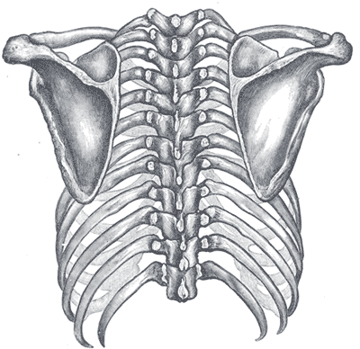
Labels on the shoulder girdle view:
- Clavicle
- Acromion
- Spine of scapula
- Scapula
On this page
What this appendicular skeleton quiz covers
The appendicular skeleton is everything that hangs from the axis of the body: the arms and legs and the two girdles that anchor them, the shoulder girdle at the top of the chest and the pelvic girdle at the hips. This quiz walks through it in six views, each on its own tab:
- Shoulder girdle: the back of the chest with both shoulder blades and collarbones. This plate has no printed labels, so the four boxes, clavicle, scapula, acromion and spine of the scapula, are placed directly on the bones.
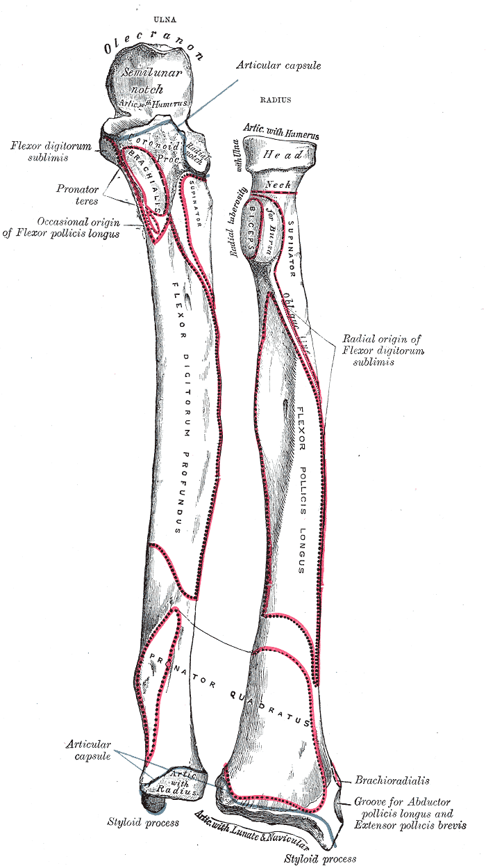
- Forearm: the ulna and radius pulled slightly apart so you can see how they fit together, from the olecranon at the elbow to the two styloid processes at the wrist.
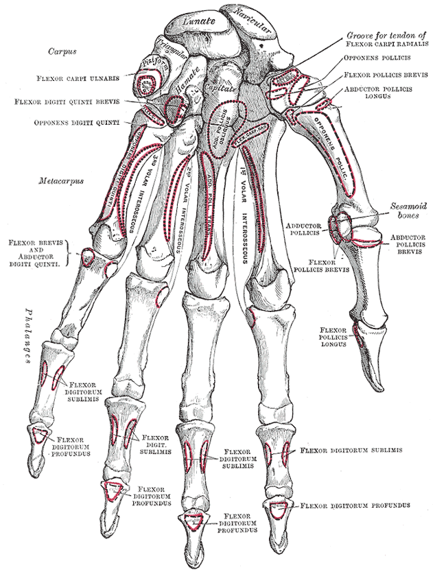
- Hand: every carpal bone of the wrist, plus the metacarpals and phalanges.
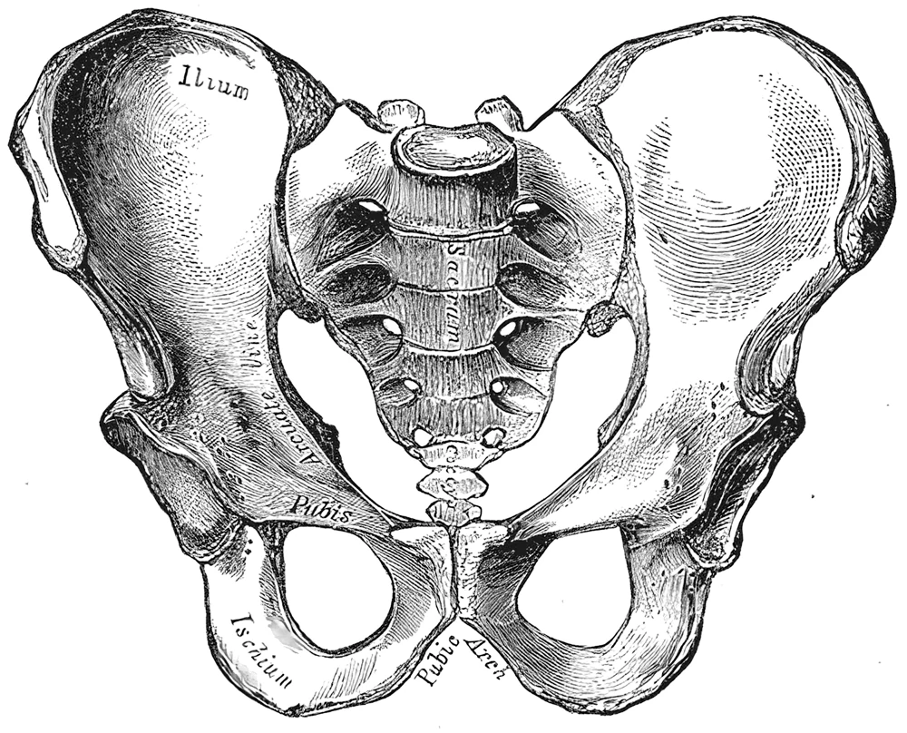
- Pelvis: the hip bones with their three parts (ilium, ischium and pubis), the pubic symphysis, the pubic arch and the obturator foramen, with the sacrum and coccyx in the middle.
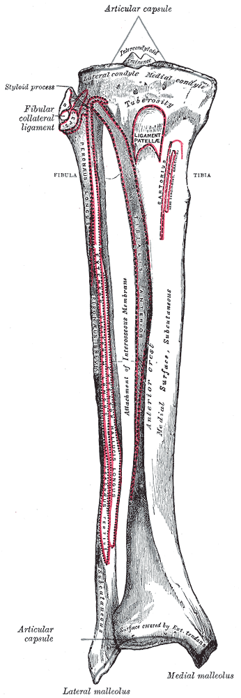
- Leg: the tibia and fibula, with the condyles and tuberosity at the knee and the two malleoli at the ankle.
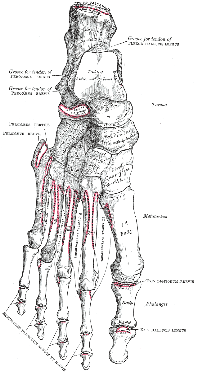
- Foot: all seven tarsal bones, plus the metatarsals and phalanges.
There are 54 labels in total. A few names on these 1918 plates have changed: the scaphoid was called the navicular, the triquetrum the triangular bone, the cuneiforms of the foot first, second and third, and the trochlear notch of the ulna the semilunar notch. The quiz asks for the current name; Type it also accepts the old one. The humerus and femur are not repeated here; you will find their landmarks in the bone markings quiz.
- Clavicle
- The collarbone: an S-shaped bone that runs from the sternum to the acromion. It is the only bony link between the arm and the trunk. (shoulder girdle view)
- Acromion
- The flat outer end of the spine of the scapula, forming the point of the shoulder. It joins the clavicle at the acromioclavicular joint. (shoulder girdle view)
- Spine of scapula
- The ridge that crosses the back of the scapula from the acromion to the medial border. (shoulder girdle view)
- Scapula
- The shoulder blade: a flat triangular bone on the back of the ribs. Together with the clavicle it forms the shoulder (pectoral) girdle. (shoulder girdle view)
- Ulna
- The forearm bone on the little-finger side. Its large upper end forms the main hinge of the elbow with the humerus. (forearm view)
- Olecranon
- The upper end of the ulna that forms the point of the elbow. The triceps attaches to it. (forearm view)
- Trochlear notch
- The deep half-moon-shaped notch at the top of the ulna that grips the trochlea of the humerus. (forearm view)
- Coronoid process
- The lip of bone at the front of the trochlear notch. It fits into the coronoid fossa of the humerus when the elbow is bent. (forearm view)
- Radial notch
- A small hollow on the side of the coronoid process where the head of the radius turns. (forearm view)
- Radius
- The forearm bone on the thumb side. Its lower end is wide and forms most of the wrist joint. (forearm view)
- Head of radius
- The disc-shaped upper end of the radius. It turns against the capitulum of the humerus and the radial notch of the ulna when you twist your forearm. (forearm view)
- Neck of radius
- The narrow part just below the head of the radius. (forearm view)
- Radial tuberosity
- A rough bump below the neck of the radius where the biceps tendon attaches. (forearm view)
- Head of ulna
- The small rounded lower end of the ulna. It joins the radius at the distal radioulnar joint, where the radius turns around it. (forearm view)
- Styloid process of ulna
- The short pointed projection at the lower end of the ulna, on the little-finger side of the wrist. (forearm view)
- Styloid process of radius
- The pointed lower end of the radius on the thumb side of the wrist. It reaches further down than the ulnar styloid. (forearm view)
- Carpal bones
- The eight small bones of the wrist, in two rows of four. (hand view)
- Scaphoid
- The boat-shaped carpal bone on the thumb side of the first row. It is the wrist bone most often broken in a fall on the outstretched hand. (hand view)
- Lunate
- The half-moon-shaped carpal bone in the middle of the first row. (hand view)
- Triquetrum
- The pyramid-shaped carpal bone on the little-finger side of the first row; the pisiform sits on its front. (hand view)
- Pisiform
- A small pea-shaped bone on the front of the triquetrum, inside the tendon of the flexor carpi ulnaris. You can feel it at the base of the palm on the little-finger side. (hand view)
- Hamate
- The carpal bone with a hook on its palm side, on the little-finger side of the second row. (hand view)
- Capitate
- The largest carpal bone, in the middle of the second row, with a rounded head that fits into the first row. (hand view)
- Trapezoid
- A small wedge-shaped carpal bone between the capitate and the trapezium; the second metacarpal sits on it. (hand view)
- Trapezium
- The carpal bone at the base of the thumb. Its saddle-shaped joint with the first metacarpal lets the thumb move across the palm. The groove for the flexor carpi radialis tendon is on it. (hand view)
- Metacarpals
- The five long bones of the palm, numbered from the thumb (first) to the little finger (fifth). (hand view)
- Phalanges of the hand
- The bones of the fingers: three in each finger (proximal, middle and distal) and two in the thumb, fourteen in all. (hand view)
- Ilium
- The large, wing-shaped upper part of the hip bone. Its upper edge is the iliac crest. (pelvis view)
- Sacrum
- The triangular bone at the back of the pelvis, made of five fused vertebrae. It joins the two hip bones at the sacroiliac joints. (pelvis view)
- Pubis
- The front part of the hip bone. The two pubic bones meet at the pubic symphysis. (pelvis view)
- Coccyx
- The tailbone, below the sacrum. (pelvis view)
- Pubic symphysis
- The joint in the midline where the two pubic bones meet, held together by a disc of fibrocartilage. (pelvis view)
- Obturator foramen
- The large opening in each hip bone below the acetabulum, framed by the pubis and the ischium. (pelvis view)
- Ischium
- The lower back part of the hip bone. Its tuberosity is the bone you sit on. (pelvis view)
- Pubic arch
- The arch formed below the pubic symphysis by the rami of the pubis and ischium on both sides. It is narrower in men, as in this plate, than in women. (pelvis view)
- Intercondylar eminence
- The raised area between the two condyles on top of the tibia; the cruciate ligaments and menisci attach around it. (leg view)
- Lateral condyle of tibia
- The outer half of the broad upper end of the tibia. The head of the fibula joins its underside. (leg view)
- Medial condyle of tibia
- The inner half of the upper end of the tibia, larger than the lateral condyle. (leg view)
- Head of fibula
- The knob at the upper end of the fibula, just below the knee on the outer side. It does not take part in the knee joint. (leg view)
- Tibial tuberosity
- The rough bump on the front of the tibia, below the knee, where the patellar ligament attaches. (leg view)
- Fibula
- The thin outer bone of the leg. It carries little weight but anchors muscles and forms the outer side of the ankle. (leg view)
- Tibia
- The shinbone: the large inner bone of the leg that carries the body's weight from the knee to the ankle. (leg view)
- Medial malleolus
- The bump on the inner side of the ankle, formed by the lower end of the tibia. (leg view)
- Lateral malleolus
- The bump on the outer side of the ankle, formed by the lower end of the fibula. It reaches lower than the medial malleolus. (leg view)
- Calcaneus
- The heel bone, the largest bone of the foot. The Achilles tendon attaches to its back. (foot view)
- Talus
- The ankle bone that sits on the calcaneus and fits between the two malleoli, carrying the weight of the body from the tibia to the foot. (foot view)
- Cuboid
- The cube-shaped tarsal bone on the outer side of the foot, in front of the calcaneus. (foot view)
- Navicular
- The boat-shaped tarsal bone on the inner side of the foot, between the talus and the three cuneiforms. (foot view)
- Lateral cuneiform
- The outermost of the three wedge-shaped cuneiform bones, next to the cuboid. (foot view)
- Intermediate cuneiform
- The smallest cuneiform, in the middle, behind the second metatarsal. (foot view)
- Medial cuneiform
- The largest cuneiform, on the inner side of the foot, behind the first metatarsal (big toe). (foot view)
- Tarsal bones
- The seven bones of the back half of the foot: calcaneus, talus, navicular, cuboid and the three cuneiforms. (foot view)
- Metatarsals
- The five long bones of the middle of the foot, numbered from the big toe (first) to the little toe (fifth). (foot view)
- Phalanges of the foot
- The bones of the toes: two in the big toe and three in each of the others. (foot view)
How to learn the appendicular skeleton
- Use the same plan for arm and leg. Each limb has one bone in the upper segment (humerus, femur), two in the lower segment (radius and ulna, tibia and fibula), a group of small bones (carpals, tarsals), five long bones (metacarpals, metatarsals) and the phalanges. Learn one limb and you have the template for the other.
- Tell the paired bones apart by side. In anatomical position the radius is on the thumb side and the ulna on the little-finger side; the tibia is on the big-toe side and the fibula on the outside. Note the difference at the ends: the thumb is lateral in the hand, but the big toe is medial in the foot.
- Learn the carpals in two rows. Both rows from the thumb side: scaphoid, lunate, triquetrum, pisiform in the first; trapezium, trapezoid, capitate, hamate in the second. To keep the two "trapez-" bones apart: the trapezium sits by the thumb.
- Build the foot from the heel. The calcaneus is at the back, the talus sits on top of it, the navicular is in front of the talus on the inner side and the cuboid in front of the calcaneus on the outer side. The three cuneiforms line up in front of the navicular.
- Feel the landmarks. The olecranon at your elbow, the styloid processes on both sides of your wrist, the malleoli on both sides of your ankle and the tibial tuberosity just below your kneecap are all easy to find on yourself.
Common mistakes
- Mixing up the forearm bones. If you hold your palm up, the radius is on the thumb side. "Radius rotates" is a good reminder: it is the bone that turns around the ulna when you flip your hand over.
- Medial and lateral malleolus. The medial malleolus belongs to the tibia, the lateral malleolus to the fibula. The lateral one reaches lower.
- Navicular in the hand and foot. In older books both a wrist bone and a foot bone are called navicular. Today the wrist bone is the scaphoid and only the foot bone keeps the name navicular.
- Putting the sacrum in the pelvic girdle. The girdle is just the two hip bones; the sacrum is part of the vertebral column.
Keep going
Take a closer look at the shoulder blade in the scapula labeling quiz, or see the whole skeleton front and back, from the skull to the feet, in the bones quiz.
Printable worksheets
Prefer paper? Each of the 6 views of this quiz comes as a free one-page PDF worksheet: the same plate with every label hidden under a blank numbered box, and a numbered line to write each name. A matching answer key lists the names with the same numbers as the quiz above.
How to use it: study the plate on screen first, then print the worksheet and fill in every line from memory. Mark it with the answer key and circle the ones you missed. A day or two later, print a fresh copy and do only the circled numbers, or switch to Type it above, which asks for the names in the same order.
- Shoulder girdle view: Worksheet · A4 · Answer key · A4
- Forearm view: Worksheet · A4 · Answer key · A4
- Hand view: Worksheet · A4 · Answer key · A4
- Pelvis view: Worksheet · A4 · Answer key · A4
- Leg view: Worksheet · A4 · Answer key · A4
- Foot view: Worksheet · A4 · Answer key · A4
Frequently asked questions
- What bones are in the appendicular skeleton?
- The bones of the upper and lower limbs and the girdles that attach them to the trunk. The pectoral (shoulder) girdle is the clavicle and scapula; the arm has the humerus, radius, ulna, carpals, metacarpals and phalanges. The pelvic girdle is the two hip bones; the leg has the femur, patella, tibia, fibula, tarsals, metatarsals and phalanges. In an adult that is 126 bones.
- Is the sacrum part of the appendicular skeleton?
- No. The sacrum and coccyx belong to the axial skeleton, as the lower end of the vertebral column. They appear in the pelvis view of this quiz because the pelvis as a whole is made of the two hip bones plus the sacrum and coccyx, but only the hip bones form the pelvic girdle.
- How can I remember the eight carpal bones?
- Learn them in two rows of four, each from the thumb side. First row: scaphoid, lunate, triquetrum, pisiform. Second row: trapezium, trapezoid, capitate, hamate. A common memory aid for that order is "Some Lovers Try Positions That They Can't Handle".
- Which forearm bone is on the thumb side?
- The radius. In anatomical position, with the palms facing forward, the radius is lateral (thumb side) and the ulna is medial (little-finger side). The ulna forms most of the elbow joint and the radius most of the wrist joint.
- What are the seven tarsal bones?
- The calcaneus (heel bone), the talus (ankle bone), the navicular, the cuboid and the three cuneiforms: medial, intermediate and lateral. Older books, including the 1918 plate in this quiz, call the cuneiforms first, second and third.
Sources
- Henry Gray, Anatomy of the Human Body, 20th ed., ed. W. H. Lewis (Philadelphia: Lea & Febiger, 1918), Osteology, Figs. 204, 213, 219, 241, 258 and 268
- Federative International Programme for Anatomical Terminology (FIPAT), Terminologia Anatomica, 2nd ed. (2019), TA2 Viewer
- OpenStax, Anatomy and Physiology 2e, chapter 8: The Appendicular Skeleton
Image credits
- Shoulder girdle view: Illustration: Henry Gray, Anatomy of the Human Body (1918), public domain, via Wikimedia Commons.
- Forearm view: Illustration: Henry Gray, Anatomy of the Human Body (1918), public domain, via Wikimedia Commons.
- Hand view: Illustration: Henry Gray, Anatomy of the Human Body (1918), public domain, via Wikimedia Commons.
- Pelvis view: Illustration: Henry Gray, Anatomy of the Human Body (1918), public domain, via Wikimedia Commons.
- Leg view: Illustration: Henry Gray, Anatomy of the Human Body (1918), public domain, via Wikimedia Commons.
- Foot view: Illustration: Henry Gray, Anatomy of the Human Body (1918), public domain, via Wikimedia Commons.
Licenses, sources and changes for every illustration are listed on the image credits page.