
Heart Labeling Quiz: Chambers, Vessels and Valves
This heart labeling quiz covers the outside of the heart, the inside of the right atrium and ventricle, and the four valves, using plates from Henry Gray's Anatomy of the Human Body (1918).



Study
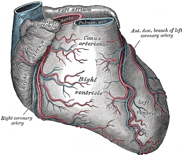
Labels on the external view:
- Left atrium
- Superior vena cava
- Aorta
- Right auricle
- Pulmonary trunk
- Anterior interventricular artery
- Conus arteriosus
- Right atrium
- Right ventricle
- Left ventricle
- Right coronary artery
On this page
What this heart labeling quiz covers
The quiz uses three plates that together show the heart from the outside in. Switch between them with the view tabs above the plate:
- External: the front (sternocostal) surface of the heart with the great vessels at the top. You label the chambers you can see from the front, the superior vena cava, aorta and pulmonary trunk, and the two coronary arteries that run over the surface.
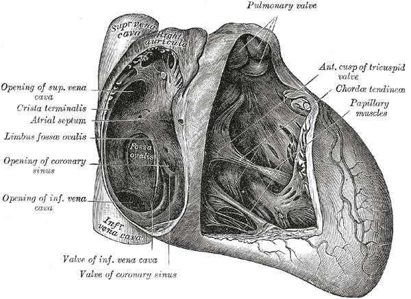
- Right side, opened: the right atrium and right ventricle cut open from the front. This is where you find the openings of the venae cavae and coronary sinus, the fossa ovalis on the wall between the atria, and the tricuspid valve with its chordae tendineae and papillary muscles.
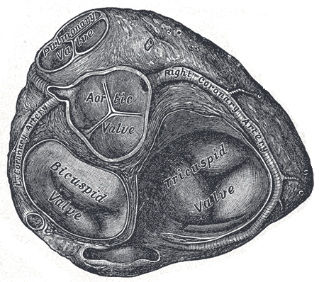
- Valves from above: the atria have been removed so you look down on the four valves in one plane, with the right and left coronary arteries curving around them.
In total there are 30 labels covering 28 different structures. Printed labels that would give away an answer or use a term no longer in use are covered; current terms that are not part of the quiz, such as the venae cavae in the opened view, are left visible.
- Left atrium
- Upper chamber on the left side of the heart. It receives oxygen-rich blood from the lungs through the pulmonary veins and passes it to the left ventricle. (external view)
- Superior vena cava
- Large vein that returns blood from the head, neck, arms and upper chest to the right atrium. (external view)
- Aorta
- The body's main artery. It leaves the left ventricle and carries oxygen-rich blood to the whole body; the coronary arteries are its first branches. (external view)
- Right auricle
- The ear-shaped pouch of the right atrium that overlaps the root of the aorta. (external view)
- Pulmonary trunk
- The large artery that leaves the right ventricle and divides into the right and left pulmonary arteries, carrying oxygen-poor blood to the lungs. (external view)
- Anterior interventricular artery
- Branch of the left coronary artery that runs down the groove between the two ventricles on the front of the heart; often called the left anterior descending (LAD) artery. (external view)
- Conus arteriosus
- The smooth-walled, funnel-shaped upper part of the right ventricle that leads into the pulmonary trunk. (external view)
- Right atrium
- Upper right chamber of the heart. It receives oxygen-poor blood from the superior and inferior venae cavae and from the coronary sinus. (external view)
- Right ventricle
- Lower right chamber. It pumps oxygen-poor blood into the pulmonary trunk and forms most of the front surface of the heart. (external view)
- Left ventricle
- Lower left chamber, with the thickest wall. It pumps oxygen-rich blood into the aorta and forms the apex of the heart. (external view)
- Right coronary artery
- Artery that runs in the groove between the right atrium and the right ventricle and supplies most of the right side of the heart. (external and valves from above view)
- Pulmonary valve
- The three-cusped valve at the exit of the right ventricle into the pulmonary trunk. It stops blood from flowing back into the ventricle. (right side, opened and valves from above view)
- Anterior cusp of tricuspid valve
- The largest of the three flaps of the tricuspid valve, which lies between the right atrium and the right ventricle. (right side, opened view)
- Opening of superior vena cava
- Where the superior vena cava empties into the upper back part of the right atrium. It has no valve. (right side, opened view)
- Chordae tendineae
- Fine tendon-like cords that tie the cusps of the atrioventricular valves to the papillary muscles and stop the cusps from turning back into the atrium. (right side, opened view)
- Papillary muscles
- Cone-shaped muscles projecting from the wall of the ventricle. They tighten the chordae tendineae when the ventricle contracts. (right side, opened view)
- Crista terminalis
- A smooth muscular ridge on the inner wall of the right atrium. It separates the smooth back part of the atrium from the rough, ridged front part. (right side, opened view)
- Interatrial septum
- The wall between the right and left atria. The fossa ovalis on its right side marks where the foramen ovale was before birth. (right side, opened view)
- Limbus of fossa ovalis
- The raised rim around the fossa ovalis on the interatrial septum. (right side, opened view)
- Fossa ovalis
- An oval hollow on the interatrial septum: the remnant of the foramen ovale, the opening that let blood bypass the lungs before birth. (right side, opened view)
- Opening of coronary sinus
- Where the coronary sinus, which collects most of the blood from the heart's own veins, drains into the right atrium, between the opening of the inferior vena cava and the tricuspid valve. (right side, opened view)
- Opening of inferior vena cava
- Where the inferior vena cava, carrying blood from the lower body, enters the lower part of the right atrium. (right side, opened view)
- Valve of inferior vena cava
- A small fold at the opening of the inferior vena cava. Before birth it helps steer blood toward the foramen ovale; it is also called the Eustachian valve. (right side, opened view)
- Valve of coronary sinus
- A thin fold that partly covers the opening of the coronary sinus; it is also called the Thebesian valve. (right side, opened view)
- Aortic valve
- The three-cusped valve between the left ventricle and the aorta. The right and left coronary arteries arise just above two of its cusps. (valves from above view)
- Left coronary artery
- Arises from the aorta just above the aortic valve and soon divides into the anterior interventricular and circumflex arteries, which supply most of the left side of the heart. (valves from above view)
- Tricuspid valve
- The valve between the right atrium and the right ventricle. It has three cusps. (valves from above view)
- Mitral valve
- The valve between the left atrium and the left ventricle. It has two cusps, which is why it is also called the bicuspid valve. (valves from above view)
How to learn the parts of the heart
- Trace the path of the blood. Start in the venae cavae, go through the right atrium and the tricuspid valve into the right ventricle, out through the pulmonary valve and trunk to the lungs, back to the left atrium, through the mitral valve into the left ventricle, and out through the aortic valve into the aorta. Once you can say this route without stopping, the labels stop being a list and become a sequence.
- Use the side as a clue. Everything on the right side of the heart handles oxygen-poor blood and leads to the lungs; everything on the left side handles oxygen-rich blood and leads to the body. Most labels in this quiz include the word right or left, so check the side first.
- Remember "tri before you bi". Blood meets the tricuspid valve (right side) before it meets the bicuspid, or mitral, valve (left side).
- Pair the two semilunar valves with their arteries. The pulmonary valve sits at the start of the pulmonary trunk and the aortic valve at the start of the aorta. In the valves view, the coronary arteries help you tell them apart: they leave the aorta just above the aortic valve.
- Study one view at a time. The external view is the easiest; the opened right side has the most small structures. Do each view in Label it before you try Type it.
Common mistakes
- Mixing up left and right on the plate. The heart is drawn as if the person faces you, so the right side of the heart is on your left. The left ventricle forms the tip (apex) on the right of the drawing.
- Atrium versus auricle. The auricle is only the small ear-shaped pouch of the atrium, not the whole chamber.
- Pulmonary trunk versus pulmonary veins. The trunk carries blood away from the right ventricle to the lungs; the pulmonary veins bring blood back from the lungs to the left atrium.
- Chordae tendineae versus papillary muscles. The papillary muscles are the muscle bumps on the ventricle wall; the chordae tendineae are the thin cords running from them to the valve cusps.
Keep going
The heart muscle itself is a special kind of muscle; compare it with the skeletal muscles of the body in the muscle quiz.
Printable worksheets
Prefer paper? Each of the 3 views of this quiz comes as a free one-page PDF worksheet: the same plate with every label hidden under a blank numbered box, and a numbered line to write each name. A matching answer key lists the names with the same numbers as the quiz above.
How to use it: study the plate on screen first, then print the worksheet and fill in every line from memory. Mark it with the answer key and circle the ones you missed. A day or two later, print a fresh copy and do only the circled numbers, or switch to Type it above, which asks for the names in the same order.
- External view: Worksheet · A4 · Answer key · A4
- Right side, opened view: Worksheet · A4 · Answer key · A4
- Valves from above view: Worksheet · A4 · Answer key · A4
Frequently asked questions
- What are the four chambers of the heart?
- Two upper chambers, the right and left atria, receive blood returning to the heart. Two lower chambers, the right and left ventricles, pump it out: the right ventricle to the lungs through the pulmonary trunk, the left ventricle to the rest of the body through the aorta.
- What are the four heart valves?
- The tricuspid valve lies between the right atrium and right ventricle, and the mitral (bicuspid) valve between the left atrium and left ventricle. The pulmonary valve guards the exit into the pulmonary trunk and the aortic valve the exit into the aorta.
- Why does the plate say bicuspid valve instead of mitral valve?
- Both names describe the same valve. Bicuspid refers to its two cusps; mitral comes from its resemblance to a bishop's mitre. The illustration dates from 1918, so the quiz uses the current name and accepts bicuspid valve when you type.
- What is the fossa ovalis?
- A shallow oval hollow on the wall between the two atria. Before birth an opening there, the foramen ovale, lets blood pass from the right atrium to the left and skip the lungs; it normally seals shortly after birth and leaves the fossa ovalis.
- Which artery is the 'widow-maker'?
- That nickname is often given to the anterior interventricular artery, also called the left anterior descending artery, because it supplies a large part of the left ventricle. In this quiz it is the vessel running down the front of the heart between the two ventricles.
Sources
- Henry Gray, Anatomy of the Human Body, 20th ed., ed. W. H. Lewis (Philadelphia: Lea & Febiger, 1918), Angiology: The Heart, Figs. 492, 493 and 495
- Federative International Programme for Anatomical Terminology (FIPAT), Terminologia Anatomica, 2nd ed. (2019), TA2 Viewer
- OpenStax, Anatomy and Physiology 2e, section 19.1: Heart Anatomy
Image credits
- External view: Illustration: Henry Gray, Anatomy of the Human Body (1918), public domain, via Wikimedia Commons.
- Right side, opened view: Illustration: Henry Gray, Anatomy of the Human Body (1918), public domain, via Wikimedia Commons.
- Valves from above view: Illustration: Henry Gray, Anatomy of the Human Body (1918), public domain, via Wikimedia Commons.
Licenses, sources and changes for every illustration are listed on the image credits page.