
Bone Markings Quiz: Humerus, Femur and Hip Bone
This bone markings quiz trains you to recognize the named bumps, ridges, holes and hollows of bones, such as tubercles, condyles, fossae and foramina, on three plates from Henry Gray's Anatomy of the Human Body (1918).



Study
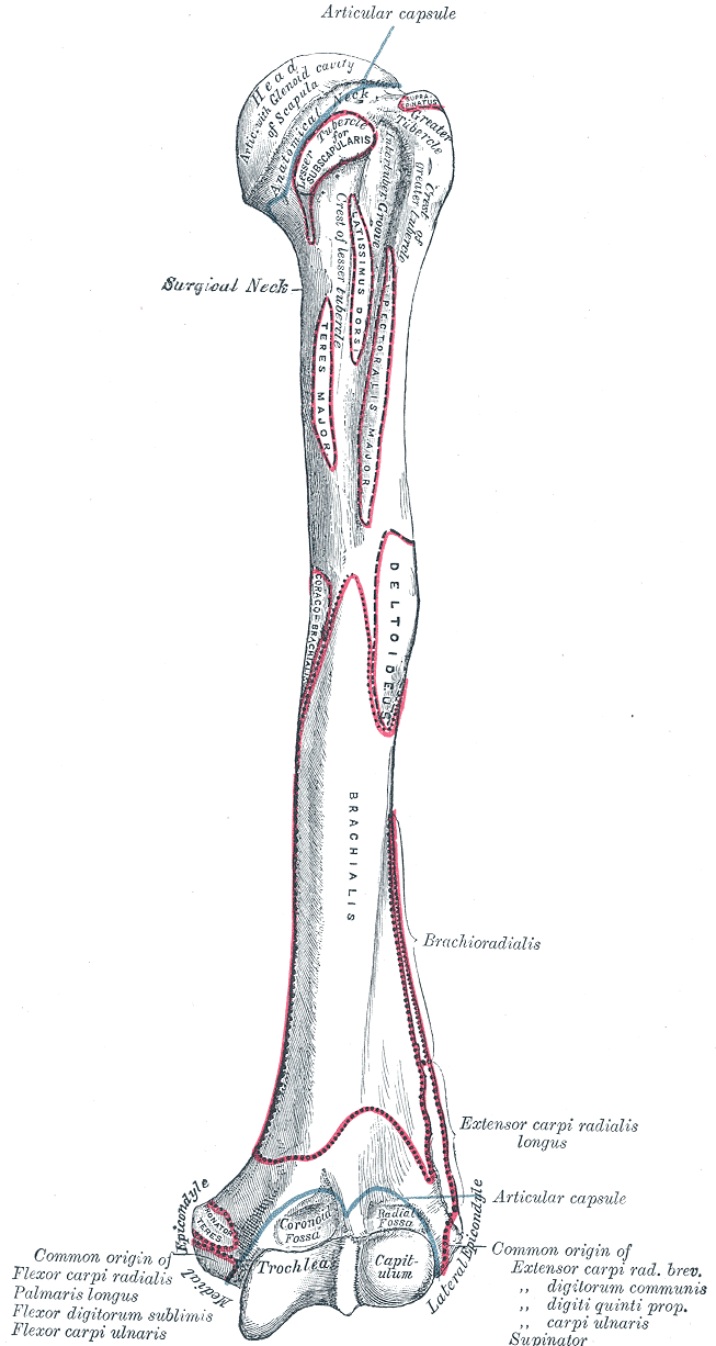
Labels on the humerus view:
- Head of humerus
- Anatomical neck
- Greater tubercle
- Lesser tubercle
- Intertubercular sulcus
- Crest of greater tubercle
- Crest of lesser tubercle
- Surgical neck
- Coronoid fossa
- Radial fossa
- Medial epicondyle of humerus
- Trochlea
- Capitulum
- Lateral epicondyle of humerus
On this page
What this bone markings quiz covers
Every bone has a vocabulary of its own, but the words repeat from bone to bone. Once you know what a tubercle, a condyle or a fossa is, you can read the name of a landmark you have never seen and guess what it looks like. This quiz trains that vocabulary on three bones, one per tab:
- Humerus: the head and the two necks, the greater and lesser tubercles with the groove and crests between them, and at the elbow end the epicondyles, the trochlea, the capitulum and two fossae.
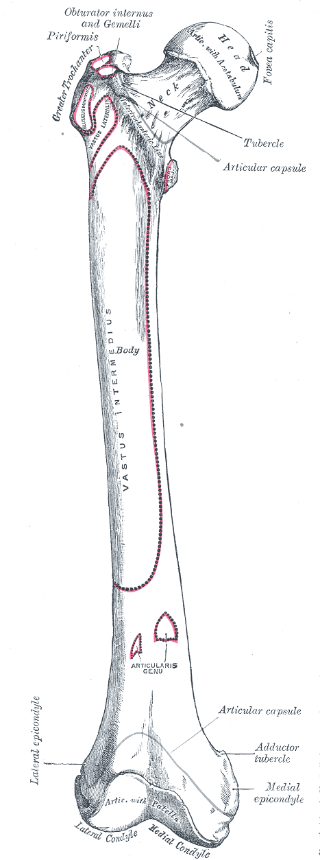
- Femur: the head with its fovea, the neck, the two trochanters and the line between them, and at the knee end the condyles, the epicondyles, the adductor tubercle and the patellar surface.
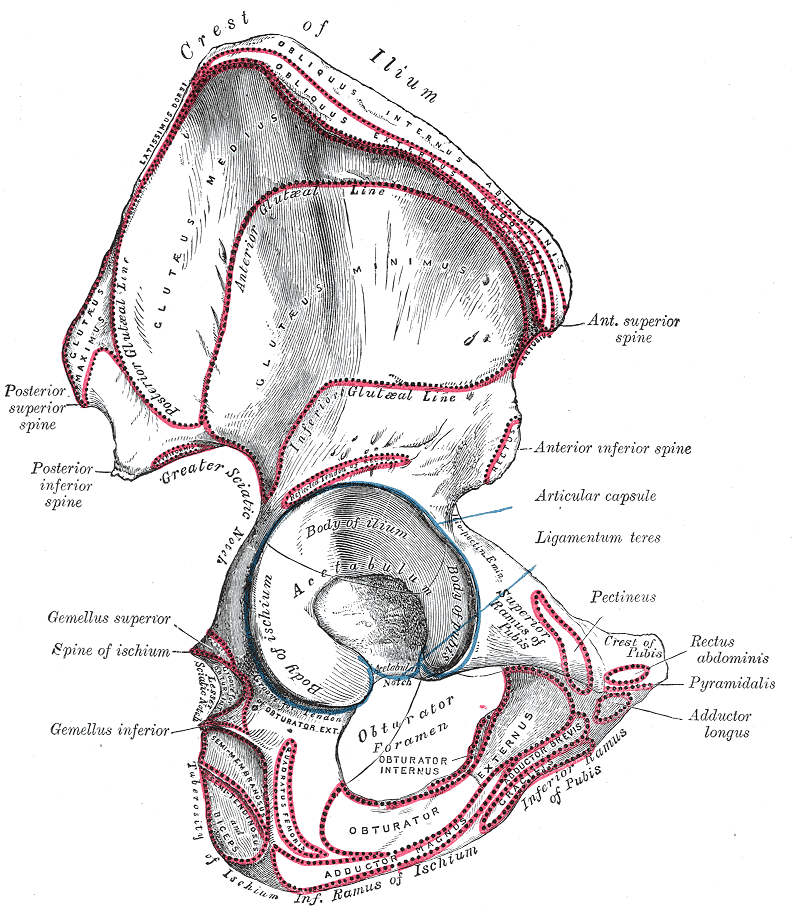
- Hip bone: the iliac crest and the four iliac spines, the acetabulum and its notch, the two sciatic notches, the ischial spine and tuberosity, the pubic crest, the three rami and the obturator foramen.
That makes 42 labels in total. The plates are from 1918, so some names are older forms, such as crest of ilium for iliac crest or intertubercular groove for intertubercular sulcus; the quiz uses the current name and Type it accepts the old one. The red outlines show where muscles attach. Their names stay visible as extra context, except a few that would give away an answer, such as the obturator muscles printed around the obturator foramen.
- Head of humerus
- A head is a rounded, smooth end of a bone that forms part of a joint. The head of the humerus fits into the glenoid cavity of the scapula at the shoulder. (humerus view)
- Anatomical neck
- A neck is the narrower part just below a head. The anatomical neck of the humerus is the groove at the edge of the joint surface, where the joint capsule attaches. (humerus view)
- Greater tubercle
- A tubercle is a small rounded bump where a muscle or ligament attaches. The greater tubercle, on the outer side of the top of the humerus, receives three of the rotator cuff muscles. (humerus view)
- Lesser tubercle
- The smaller bump on the front of the top of the humerus. The subscapularis muscle attaches to it. (humerus view)
- Intertubercular sulcus
- A sulcus or groove is a long, narrow channel in a bone. The intertubercular sulcus runs between the two tubercles and carries the tendon of the long head of the biceps. (humerus view)
- Crest of greater tubercle
- A crest is a prominent ridge. This one runs down from the greater tubercle along the outer edge of the groove; the pectoralis major attaches to it. (humerus view)
- Crest of lesser tubercle
- The ridge that runs down from the lesser tubercle along the inner edge of the groove; the teres major attaches to it. (humerus view)
- Surgical neck
- The narrowing of the shaft just below the tubercles. It is named “surgical” because it is a common place for fractures of the humerus. (humerus view)
- Coronoid fossa
- A fossa is a shallow hollow in a bone. The coronoid fossa, on the front just above the trochlea, receives the coronoid process of the ulna when the elbow is fully bent. (humerus view)
- Radial fossa
- A small hollow above the capitulum that receives the edge of the head of the radius when the elbow is fully bent. (humerus view)
- Medial epicondyle of humerus
- An epicondyle is a bump just above a condyle. The medial epicondyle is the large one on the inner side of the elbow; the ulnar nerve runs behind it (the “funny bone”). (humerus view)
- Trochlea
- A trochlea is a pulley-shaped joint surface. The trochlea of the humerus joins the ulna at the elbow. (humerus view)
- Capitulum
- A small, rounded knob (literally “little head”) on the outer part of the lower end of the humerus. It joins the head of the radius. (humerus view)
- Lateral epicondyle of humerus
- The smaller bump on the outer side of the elbow. Most of the muscles that extend the wrist and fingers arise from it. (humerus view)
- Head of femur
- The ball-shaped upper end of the femur. It fits into the acetabulum of the hip bone to form the hip joint. (femur view)
- Fovea capitis
- A fovea is a small pit. The fovea capitis is the pit near the center of the femoral head where the ligament of the head of the femur (ligamentum teres) attaches. (femur view)
- Neck of femur
- The narrow part that joins the head to the shaft at an angle. It is the usual site of hip fractures in older people. (femur view)
- Greater trochanter
- A trochanter is a very large, blunt process; only the femur has them. The greater trochanter is the bump you can feel on the side of the hip, where the gluteus medius and minimus attach. (femur view)
- Intertrochanteric line
- A line is a low, narrow ridge, smaller than a crest. The intertrochanteric line runs between the two trochanters on the front of the femur; the hip joint capsule attaches to it. (femur view)
- Lesser trochanter
- The smaller, cone-shaped process on the inner side of the femur below the neck. The iliopsoas, the strongest hip flexor, attaches to it. (femur view)
- Lateral epicondyle of femur
- The bump on the outer side of the lower end of the femur, above the lateral condyle. The fibular collateral ligament of the knee attaches to it. (femur view)
- Adductor tubercle
- A small bump at the top of the medial epicondyle where the tendon of the adductor magnus attaches. (femur view)
- Medial epicondyle of femur
- The bump on the inner side of the lower end of the femur, above the medial condyle. The tibial collateral ligament of the knee attaches to it. (femur view)
- Patellar surface
- The smooth, grooved joint surface on the front of the lower femur where the kneecap glides. (femur view)
- Lateral condyle of femur
- A condyle is a large, rounded joint surface at the end of a bone. The two femoral condyles rest on the tibia at the knee. (femur view)
- Medial condyle of femur
- The inner of the two rounded knuckles at the lower end of the femur. It reaches further down than the lateral condyle. (femur view)
- Iliac crest
- The long curved upper border of the ilium, the part you feel when you put your hands on your hips. The abdominal wall muscles attach along it. (hip bone view)
- Anterior superior iliac spine
- A spine is a sharp, slender projection. The anterior superior iliac spine is the front end of the iliac crest, an easy landmark to feel at the front of the hip; the sartorius arises from it. (hip bone view)
- Posterior superior iliac spine
- The back end of the iliac crest. It lies under the small dimples you can see on the lower back on each side of the spine. (hip bone view)
- Anterior inferior iliac spine
- The smaller projection below the anterior superior iliac spine, above the acetabulum. The straight head of the rectus femoris arises from it. (hip bone view)
- Posterior inferior iliac spine
- The projection below the posterior superior iliac spine, at the top of the greater sciatic notch. (hip bone view)
- Greater sciatic notch
- A notch is an indentation at the edge of a bone. The greater sciatic notch is a deep curve on the back of the hip bone; the sciatic nerve and the piriformis pass through it. (hip bone view)
- Acetabulum
- The deep cup-shaped socket on the outer side of the hip bone that holds the head of the femur. All three bones of the hip, ilium, ischium and pubis, meet in it. (hip bone view)
- Ischial spine
- A pointed projection on the back of the ischium, between the greater and lesser sciatic notches. The sacrospinous ligament attaches to it. (hip bone view)
- Acetabular notch
- The gap in the lower rim of the acetabulum, bridged by a ligament. Blood vessels to the head of the femur pass through it. (hip bone view)
- Lesser sciatic notch
- The smaller notch below the ischial spine. The tendon of the obturator internus turns around it. (hip bone view)
- Superior pubic ramus
- A ramus is a branch-like bar of bone. The superior pubic ramus runs from the body of the pubis to the acetabulum and forms the upper edge of the obturator foramen. (hip bone view)
- Pubic crest
- The short rough ridge on the upper edge of the body of the pubis, near the midline. The rectus abdominis attaches to it. (hip bone view)
- Obturator foramen
- A foramen is a hole through a bone. The obturator foramen is the large opening below the acetabulum; in life it is almost closed by a membrane, leaving a small canal for the obturator nerve and vessels. (hip bone view)
- Inferior pubic ramus
- The bar of bone that runs down and back from the body of the pubis to join the ramus of the ischium. (hip bone view)
- Ischial tuberosity
- A tuberosity is a large, rough bump. The ischial tuberosity carries your weight when you sit, and the hamstring muscles arise from it. (hip bone view)
- Ischial ramus
- The bar of bone that runs forward from the ischial tuberosity to meet the inferior pubic ramus, closing the obturator foramen from below. (hip bone view)
The main types of bone markings
| Type | What it is | Example in this quiz |
|---|---|---|
| Head | Rounded joint end of a bone, on a neck | Head of femur |
| Condyle | Large rounded joint surface | Medial condyle of femur |
| Trochlea | Pulley-shaped joint surface | Trochlea of humerus |
| Epicondyle | Bump above a condyle | Lateral epicondyle of humerus |
| Tubercle | Small rounded bump | Greater tubercle |
| Tuberosity | Large rough bump | Ischial tuberosity |
| Trochanter | Very large blunt process (femur only) | Greater trochanter |
| Spine | Sharp, slender projection | Anterior superior iliac spine |
| Crest / line | Prominent ridge / low ridge | Iliac crest, intertrochanteric line |
| Fossa | Shallow hollow | Coronoid fossa |
| Sulcus | Groove | Intertubercular sulcus |
| Notch | Indentation in an edge | Greater sciatic notch |
| Foramen | Hole through a bone | Obturator foramen |
| Ramus | Bar-like branch of a bone | Superior pubic ramus |
How to learn bone markings
- Learn the type first, then the bone. Before each round, read the table above. When you meet a new name, split it into type and place: "lateral epicondyle" is an epicondyle on the outer side.
- Ask what each marking is for. Projections such as tubercles, crests and spines are where muscles and ligaments pull on the bone. Smooth heads, condyles and the trochlea form joints. Holes and grooves let something pass: the biceps tendon in the intertubercular sulcus, nerves and vessels through the obturator foramen.
- Compare the arm and the leg. The humerus and femur share a plan: a head and neck at the top, bumps for muscles below the head, condyles and epicondyles at the lower end. Do the two tabs back to back and the pattern sticks.
- Feel them on yourself. You can feel the medial epicondyle of the humerus at your elbow, the greater trochanter on the side of your hip, the iliac crest and anterior superior iliac spine at your waist, and the ischial tuberosities when you sit on a hard chair.
Common mistakes
- Medial and lateral epicondyles of the humerus. The plate shows a left humerus from the front, so the medial epicondyle, the larger one, is on the left of the drawing.
- Coronoid fossa versus radial fossa. Both are above the elbow joint on the front. The coronoid fossa sits above the trochlea and fits the ulna; the radial fossa sits above the capitulum and fits the radius.
- Anterior superior versus anterior inferior iliac spine. The superior one is the front tip of the iliac crest; the inferior one is lower down, just above the acetabulum.
- Greater versus lesser sciatic notch. The greater notch is above the ischial spine and much bigger; the lesser notch is below it.
Keep going
Bone markings are named on every bone of the limbs. See where these bones sit in the body, together with the scapula, clavicle and the bones of the hands and feet, in the appendicular skeleton quiz, or label the borders and fossae of the shoulder blade in the scapula labeling quiz.
Printable worksheets
Prefer paper? Each of the 3 views of this quiz comes as a free one-page PDF worksheet: the same plate with every label hidden under a blank numbered box, and a numbered line to write each name. A matching answer key lists the names with the same numbers as the quiz above.
How to use it: study the plate on screen first, then print the worksheet and fill in every line from memory. Mark it with the answer key and circle the ones you missed. A day or two later, print a fresh copy and do only the circled numbers, or switch to Type it above, which asks for the names in the same order.
- Humerus view: Worksheet · A4 · Answer key · A4
- Femur view: Worksheet · A4 · Answer key · A4
- Hip bone view: Worksheet · A4 · Answer key · A4
Frequently asked questions
- What are bone markings?
- Bone markings are the named features on the surface of a bone: bumps and ridges where muscles and ligaments attach, smooth rounded ends that form joints, and holes, grooves and hollows that let nerves and blood vessels pass or that hold other structures. Each type has its own name, such as tubercle, condyle, fossa or foramen.
- What is the difference between a tubercle, a tuberosity and a trochanter?
- All three are rough projections where muscles attach; they differ in size. A tubercle is small and rounded, like the greater tubercle of the humerus. A tuberosity is larger and rougher, like the ischial tuberosity you sit on. A trochanter is a very large, blunt process, and the only ones in the body are on the femur.
- What is the difference between a condyle and an epicondyle?
- A condyle is a large, smooth, rounded joint surface at the end of a bone, like the condyles of the femur that rest on the tibia. An epicondyle is a smaller rough bump just above a condyle, outside the joint, where ligaments and muscles attach.
- What is the difference between a fossa and a foramen?
- A fossa is a shallow hollow on the surface of a bone, such as the coronoid fossa of the humerus. A foramen goes all the way through the bone, like the obturator foramen of the hip bone.
- Why does the humerus have two necks?
- The anatomical neck is the narrow groove right at the edge of the joint surface of the head. The surgical neck is the narrowing of the shaft just below the tubercles; it gets its name because fractures happen there far more often, so surgeons deal with it more.
Sources
- Henry Gray, Anatomy of the Human Body, 20th ed., ed. W. H. Lewis (Philadelphia: Lea & Febiger, 1918), Osteology, Figs. 207, 235 and 244
- Federative International Programme for Anatomical Terminology (FIPAT), Terminologia Anatomica, 2nd ed. (2019), TA2 Viewer
- OpenStax, Anatomy and Physiology 2e, section 6.3: Bone Structure
Image credits
- Humerus view: Illustration: Henry Gray, Anatomy of the Human Body (1918), public domain, via Wikimedia Commons.
- Femur view: Illustration: Henry Gray, Anatomy of the Human Body (1918), public domain, via Wikimedia Commons.
- Hip bone view: Illustration: Henry Gray, Anatomy of the Human Body (1918), public domain, via Wikimedia Commons.
Licenses, sources and changes for every illustration are listed on the image credits page.