
Muscle Quiz: Label the Superficial Muscles of the Body
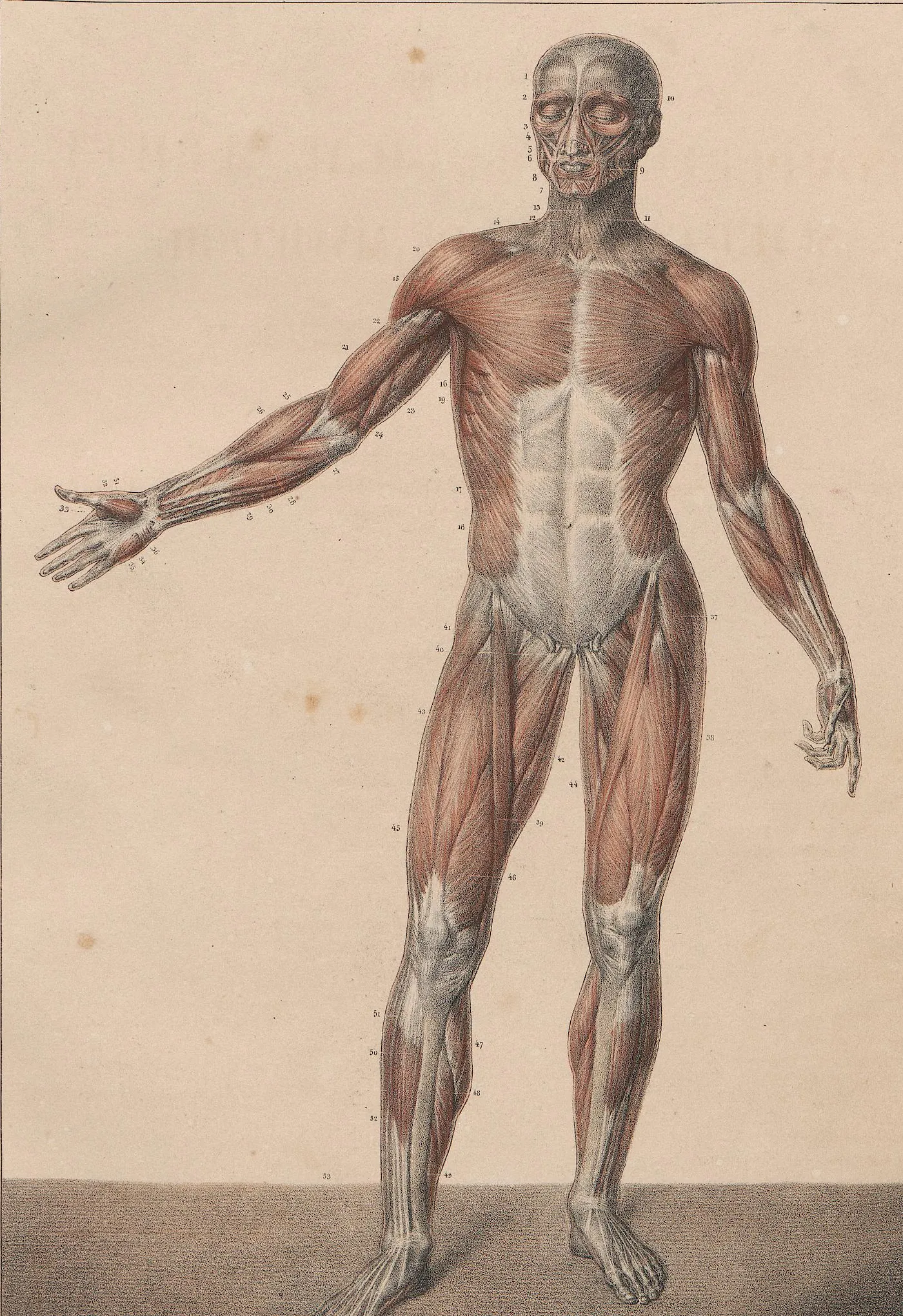
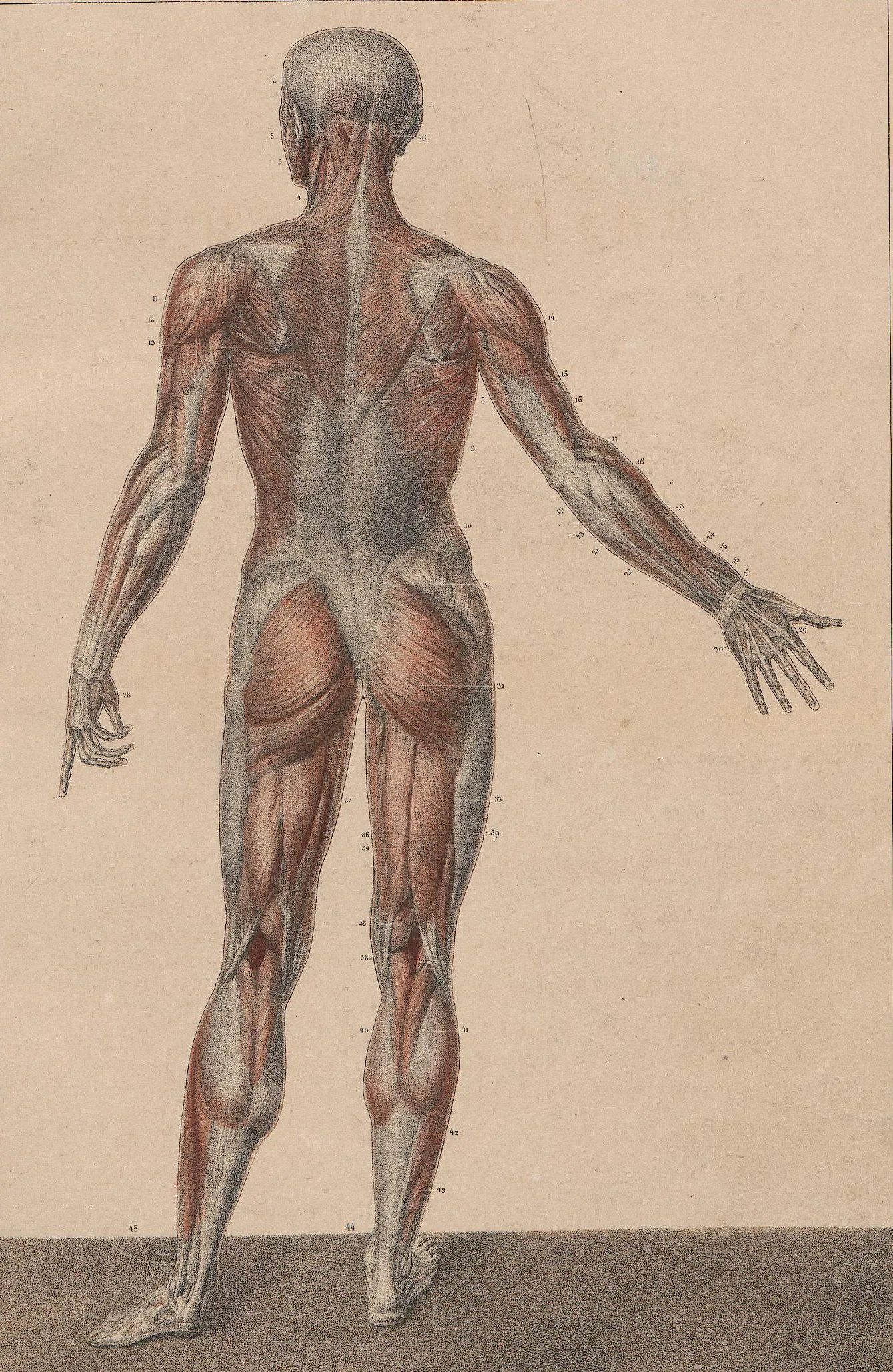
This muscle quiz asks you to label the main superficial muscles of the human body, from the face to the legs, on a front and a back view from Bourgery's anatomical atlas (1831).
Study
Labels on the anterior (front) view:
- Frontalis
- Orbicularis oculi
- Temporalis
- Zygomaticus major
- Orbicularis oris
- Masseter
- Sternocleidomastoid
- Trapezius
- Deltoid
- Pectoralis major
- Biceps brachii
- Serratus anterior
- Brachioradialis
- Latissimus dorsi
- Triceps brachii
- Brachialis
- Pronator teres
- Flexor carpi radialis
- External oblique
- Flexor carpi ulnaris
- Palmaris longus
- Rectus abdominis
- Tensor fasciae latae
- Iliopsoas
- Rectus femoris
- Iliotibial tract
- Adductor longus
- Gracilis
- Sartorius
- Vastus lateralis
- Vastus medialis
- Extensor digitorum longus
- Gastrocnemius
- Tibialis anterior
- Soleus
- Fibularis longus
On this page
What this muscle quiz covers
This quiz covers the superficial muscles of the whole body, the layer you see first under the skin, on two full-length plates. Switch between them with the tabs above the plate:
- Anterior (front): the muscles of the face and neck, the chest and abdomen (pectoralis major, serratus anterior, external oblique, rectus abdominis), the shoulder and the front of the arm and forearm, and the hip, thigh and leg muscles seen from the front, including the quadriceps, the adductors and the sartorius.
- Posterior (back): the muscles of the back of the neck and the back (trapezius, latissimus dorsi, rhomboid, the rotator cuff muscles on the shoulder blade), the triceps and the extensors on the back of the forearm, the gluteal muscles, the hamstrings and the calf.
That is 61 labels in total. A few muscles show up in both views, such as the trapezius, deltoid, latissimus dorsi, external oblique, gastrocnemius and soleus; seeing them from both sides is the best way to understand their shape. The plates come from Bourgery and Jacob's anatomical atlas of 1831. Bourgery marks each muscle with a small number and names it on a separate legend page in French, sometimes with older names such as "long supinateur" for the brachioradialis. The quiz uses the current English names; each box sits on Bourgery's number for that muscle, and the numbers not used in the quiz stay as printed.
- Frontalis
- The thin muscle of the forehead, the front part of the occipitofrontalis. It raises the eyebrows and wrinkles the forehead. (anterior (front) view)
- Orbicularis oculi
- The ring of muscle around the eye. It closes the eyelids, gently when you blink and tightly when you squeeze. (anterior (front) view)
- Temporalis
- A fan-shaped chewing muscle on the side of the head that closes the jaw. (anterior (front) view)
- Zygomaticus major
- Runs from the cheekbone to the corner of the mouth and pulls it up and back when you smile. (anterior (front) view)
- Orbicularis oris
- The ring of muscle in the lips. It closes and purses the lips. (anterior (front) view)
- Masseter
- The thick chewing muscle at the angle of the jaw; you can feel it tighten when you clench your teeth. (anterior (front) view)
- Sternocleidomastoid
- The long muscle on each side of the neck, from the sternum and clavicle to the mastoid process behind the ear. It turns and bends the head. (anterior (front) view)
- Trapezius
- The large diamond-shaped muscle of the upper back and neck. It lifts, pulls back and rotates the shoulder blade.
- Deltoid
- The rounded muscle that caps the shoulder. It lifts the arm away from the body.
- Pectoralis major
- The large fan-shaped chest muscle. It pulls the arm across the chest and rotates it inward. (anterior (front) view)
- Biceps brachii
- The two-headed muscle on the front of the upper arm. It bends the elbow and turns the palm up. (anterior (front) view)
- Serratus anterior
- A saw-toothed muscle on the side of the chest that holds the shoulder blade against the ribs and pulls it forward, as in a punch. (anterior (front) view)
- Brachioradialis
- The muscle that forms the bulge on the thumb side of the forearm, just below the elbow. It bends the elbow. (anterior (front) view)
- Latissimus dorsi
- The broad muscle of the lower back and side. It pulls the arm down and back, as in a pull-up or a swimming stroke.
- Triceps brachii
- The three-headed muscle on the back of the upper arm. It straightens the elbow.
- Brachialis
- Lies under the biceps on the front of the upper arm; the main muscle that bends the elbow. (anterior (front) view)
- Pronator teres
- A short muscle across the front of the upper forearm that turns the palm down. (anterior (front) view)
- Flexor carpi radialis
- A forearm muscle that bends the wrist and tilts it toward the thumb side. (anterior (front) view)
- External oblique
- The outermost muscle on the side of the abdomen. It bends and twists the trunk.
- Flexor carpi ulnaris
- The forearm muscle on the little-finger side that bends the wrist and tilts it toward the little finger. (anterior (front) view)
- Palmaris longus
- A slender forearm muscle with a long tendon to the palm. It is missing in some people. (anterior (front) view)
- Rectus abdominis
- The long, flat muscle down the front of the abdomen, crossed by tendinous bands that give the “six-pack” look. It bends the trunk forward. (anterior (front) view)
- Tensor fasciae latae
- A short muscle at the front of the hip that tightens the iliotibial tract and helps flex and abduct the thigh. (anterior (front) view)
- Iliopsoas
- The combined psoas major and iliacus, the strongest hip flexor. In this view only its lower end is seen, in the groin. (anterior (front) view)
- Rectus femoris
- The part of the quadriceps that runs straight down the front of the thigh. It straightens the knee and flexes the hip. (anterior (front) view)
- Iliotibial tract
- A thick band of fascia down the outer side of the thigh, from the hip to below the knee. (anterior (front) view)
- Adductor longus
- A muscle on the inner thigh that pulls the leg toward the midline. (anterior (front) view)
- Gracilis
- A long, thin muscle on the inner side of the thigh that pulls the leg inward and bends the knee. (anterior (front) view)
- Sartorius
- The longest muscle in the body, running diagonally across the front of the thigh. It helps you sit cross-legged. (anterior (front) view)
- Vastus lateralis
- The large outer part of the quadriceps, on the side of the thigh. (anterior (front) view)
- Vastus medialis
- The inner part of the quadriceps, forming the bulge above the inner side of the knee. (anterior (front) view)
- Extensor digitorum longus
- A muscle on the front of the leg, beside the tibialis anterior, that straightens the four lesser toes. (anterior (front) view)
- Gastrocnemius
- The two-headed calf muscle. It points the foot and helps bend the knee.
- Tibialis anterior
- The muscle along the front of the shin that lifts the foot. (anterior (front) view)
- Soleus
- A flat muscle under the gastrocnemius. Together they form the calf and share the Achilles tendon.
- Fibularis longus
- A muscle on the outer side of the leg whose tendon passes behind the ankle and under the foot. It points the foot and turns the sole outward. (anterior (front) view)
- Occipitalis
- The back part of the occipitofrontalis, over the back of the skull. It pulls the scalp backward. (posterior (back) view)
- Splenius capitis
- A strap-like muscle in the back of the neck, under the trapezius, that extends and turns the head. (posterior (back) view)
- Infraspinatus
- A rotator cuff muscle in the infraspinous fossa of the scapula. It turns the arm outward. (posterior (back) view)
- Teres minor
- A small rotator cuff muscle along the lateral border of the scapula, below the infraspinatus. It turns the arm outward. (posterior (back) view)
- Teres major
- A thick muscle from the lower angle of the scapula to the humerus. It pulls the arm down and back. (posterior (back) view)
- Rhomboid major
- Runs from the spine to the medial border of the shoulder blade and pulls the shoulder blades together. (posterior (back) view)
- Extensor carpi radialis longus
- A muscle on the back of the forearm, thumb side, that straightens the wrist and tilts it toward the thumb. (posterior (back) view)
- Extensor carpi radialis brevis
- Lies just beside the longus and also straightens the wrist. (posterior (back) view)
- Anconeus
- A small triangular muscle at the back of the elbow that helps the triceps straighten it. (posterior (back) view)
- Extensor carpi ulnaris
- A muscle on the little-finger side of the back of the forearm that straightens the wrist and tilts it toward the little finger. (posterior (back) view)
- Extensor digitorum
- The main muscle on the back of the forearm that straightens the four fingers. (posterior (back) view)
- Extensor digiti minimi
- A thin muscle beside the extensor digitorum that straightens the little finger. (posterior (back) view)
- Gluteus medius
- A muscle on the outer side of the hip, above the gluteus maximus, that keeps the pelvis level when you walk. (posterior (back) view)
- Gluteus maximus
- The largest muscle of the buttock. It straightens the hip, as when you climb stairs or stand up. (posterior (back) view)
- Biceps femoris
- The outer hamstring muscle at the back of the thigh. It bends the knee and straightens the hip. (posterior (back) view)
- Adductor magnus
- The largest adductor muscle, on the inner back of the thigh. (posterior (back) view)
- Semitendinosus
- An inner hamstring muscle with a long tendon at its lower end. (posterior (back) view)
- Semimembranosus
- The deepest inner hamstring muscle, under the semitendinosus. (posterior (back) view)
How to learn the muscles
- Decode the names. Most muscle names describe the muscle: biceps has two heads, triceps three; rectus means straight; maximus, medius and minimus give the size; flexor and extensor give the action. If you know the Latin pieces, half the work is done.
- Learn muscles in groups that work together. Rotator cuff: supraspinatus, infraspinatus, teres minor and subscapularis. Quadriceps: rectus femoris and the three vasti. Hamstrings: biceps femoris, semitendinosus and semimembranosus. Calf: gastrocnemius and soleus.
- Pair front and back. Biceps and triceps, quadriceps and hamstrings, tibialis anterior and the calf muscles: each pair pulls in opposite directions across the same joint.
- Move while you study. Bend your elbow and feel the biceps tighten; shrug and feel the trapezius; stand on tiptoe and feel the calf. Linking a name to a movement makes it much easier to remember.
- Start with Study mode on one view. Read each description, then switch to Label it. The front view has more labels, so many students find the back view a good place to start.
Common mistakes
- Deltoid versus trapezius. The deltoid caps the shoulder; the trapezius covers the upper back and neck and reaches the shoulder only at its outer edge.
- Biceps brachii versus biceps femoris. Brachii means of the arm, femoris of the thigh. The biceps femoris is a hamstring, at the back of the thigh.
- Sartorius versus rectus femoris. The sartorius crosses the thigh diagonally from the outer hip to the inner knee; the rectus femoris runs straight down the middle.
- Gastrocnemius versus soleus. The gastrocnemius forms the two rounded bulges of the calf; the soleus lies underneath and shows at the sides, lower down.
- Latissimus dorsi in the front view. Only its edge shows at the side of the chest from the front; in the back view it covers most of the lower back.
Keep going
The heart is a muscle too, but a very different kind; see its chambers and valves in the heart labeling quiz. If terms like brachial, femoral or popliteal are still new, the regional terms anatomy quiz covers the names of the body regions these muscles sit in.
Printable worksheets
Prefer paper? Each of the 2 views of this quiz comes as a free one-page PDF worksheet: the same plate with every label hidden under a blank numbered box, and a numbered line to write each name. A matching answer key lists the names with the same numbers as the quiz above.
How to use it: study the plate on screen first, then print the worksheet and fill in every line from memory. Mark it with the answer key and circle the ones you missed. A day or two later, print a fresh copy and do only the circled numbers, or switch to Type it above, which asks for the names in the same order.
- Anterior (front) view: Worksheet · A4 · Answer key · A4
- Posterior (back) view: Worksheet · A4 · Answer key · A4
Frequently asked questions
- How many muscles are in the human body?
- Around 600 skeletal muscles, depending on how they are counted (some muscles have several parts that may be counted separately). This quiz covers the main superficial ones, the muscles you would see first if the skin were removed.
- What is the largest muscle in the body?
- The gluteus maximus, the main muscle of the buttock, is usually named the largest by mass. The longest is the sartorius, which runs diagonally across the front of the thigh. Both are in this quiz.
- Which muscles make up the quadriceps and the hamstrings?
- The quadriceps, on the front of the thigh, are the rectus femoris, vastus lateralis, vastus medialis and vastus intermedius (the last one lies deep and is not visible here). The hamstrings, on the back of the thigh, are the biceps femoris, semitendinosus and semimembranosus.
- How are muscles named?
- Muscle names describe something about the muscle: its shape (deltoid, trapezius), size (gluteus maximus, teres minor), location (tibialis anterior, temporalis), number of heads (biceps, triceps), direction of fibers (rectus, oblique), attachments (sternocleidomastoid) or action (flexor, extensor, adductor). Reading a name this way often tells you where to look.
- Is the figure in this quiz nude?
- The figure is an anatomical drawing from 1831 with the skin removed to show the muscles. The genitals were left out of the original engraving, and the plates are shown as published, without any other change.
Sources
- Jean-Baptiste Marc Bourgery and Nicolas-Henri Jacob, Traité complet de l'anatomie de l'homme, Atlas II (Paris, 1831), Pl. 60 and 61 with their legends; digitization: Heidelberg University Library
- Federative International Programme for Anatomical Terminology (FIPAT), Terminologia Anatomica, 2nd ed. (2019), TA2 Viewer
- OpenStax, Anatomy and Physiology 2e, chapter 11: The Muscular System
Image credits
- Anterior (front) view: Illustration: J.-M. Bourgery and N.-H. Jacob, Traité complet de l'anatomie de l'homme (1831–1854), public domain, via Wikimedia Commons. Digitization: Heidelberg University Library.
- Posterior (back) view: Illustration: J.-M. Bourgery and N.-H. Jacob, Traité complet de l'anatomie de l'homme (1831–1854), public domain, via Wikimedia Commons. Digitization: Heidelberg University Library.
Licenses, sources and changes for every illustration are listed on the image credits page.