
Eye Anatomy Quiz: Parts of the Eye in Cross-Section
This eye anatomy quiz uses a horizontal section of the eyeball from Henry Gray's Anatomy of the Human Body (1918) to test the layers, chambers and inner structures of the eye.
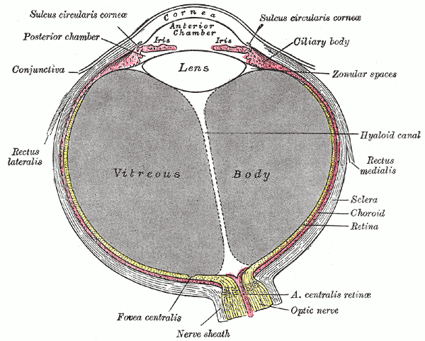

Study
Labels on the horizontal section view:
- Cornea
- Anterior chamber
- Posterior chamber
- Iris
- Ciliary body
- Lens
- Conjunctiva
- Hyaloid canal
- Lateral rectus
- Medial rectus
- Vitreous body
- Sclera
- Choroid
- Retina
- Central retinal artery
- Optic nerve
- Fovea centralis
On this page
What this eye anatomy quiz covers
The quiz is built on a horizontal section of the eyeball: imagine cutting the eye straight across at the level of the pupil and looking down on the cut surface. The cornea is at the top of the drawing and the optic nerve leaves at the bottom. The plate shows all the main parts of the eye in a single view, from the clear front surface to the layers that line the back of the eye, plus two of the muscles that move it.
There are 17 labels to learn. They fall into four groups:
- The outer coat: cornea and sclera, plus the conjunctiva that covers the front of the sclera.
- The middle, vascular coat: choroid, ciliary body and iris.
- The inner coat and the light path: retina, fovea centralis, optic nerve and the central retinal artery.
- The contents of the eyeball: anterior and posterior chambers, lens, vitreous body and the hyaloid canal that runs through it, with the lateral and medial rectus muscles on each side.
Two printed labels on the original plate use an old Latin term, sulcus circularis corneae (the groove where the cornea meets the sclera), and are covered; the other printed labels that are not part of the quiz, such as the nerve sheath, are left visible.
- Cornea
- The clear, curved front of the eye. It does most of the focusing of incoming light and has no blood vessels.
- Anterior chamber
- The space between the cornea and the iris. It is filled with aqueous humor, which nourishes the cornea and the lens.
- Posterior chamber
- The narrow space behind the iris and in front of the lens. Aqueous humor made by the ciliary body collects here and flows through the pupil into the anterior chamber.
- Iris
- The colored ring of muscle around the pupil. It controls how much light enters the eye by changing the size of the pupil.
- Ciliary body
- A ring of muscle and folds just behind the iris. It produces aqueous humor and changes the shape of the lens to focus on near objects.
- Lens
- A clear, flexible disk behind the iris. It fine-tunes focus by changing shape and is held in place by the fibers of the ciliary zonule.
- Conjunctiva
- The thin, transparent membrane that covers the white of the eye and lines the inside of the eyelids.
- Hyaloid canal
- A narrow channel through the vitreous body, from the optic disc to the back of the lens. It is what remains of the hyaloid artery, which supplies the lens before birth.
- Lateral rectus
- One of the six muscles that move the eye. It turns the eye outward, away from the nose, and is supplied by the abducens nerve (cranial nerve VI).
- Medial rectus
- The eye muscle that turns the eye inward, toward the nose. It is supplied by the oculomotor nerve (cranial nerve III).
- Vitreous body
- The clear gel that fills the large space behind the lens and helps hold the retina in place.
- Sclera
- The tough, white outer coat of the eyeball. It protects the eye and gives the eye muscles a firm attachment.
- Choroid
- The thin layer rich in blood vessels between the sclera and the retina. It nourishes the outer layers of the retina.
- Retina
- The light-sensitive inner layer of the eye. Its rods and cones turn light into nerve signals that leave the eye through the optic nerve.
- Central retinal artery
- A branch of the ophthalmic artery that runs inside the optic nerve and supplies the inner layers of the retina.
- Optic nerve
- Cranial nerve II. It carries visual signals from the retina to the brain.
- Fovea centralis
- A small pit in the center of the macula, packed with cones. It gives the sharpest central vision.
How to remember the parts of the eye
- Think of three coats, like an onion. The tough outer coat (sclera and cornea), the blood-rich middle coat (choroid, ciliary body and iris) and the nervous inner coat (retina). Every label on the plate except the contents fits one of these three.
- Follow a ray of light. Light passes through the cornea, the aqueous humor of the anterior chamber, the pupil framed by the iris, the lens, the vitreous body and finally reaches the retina, with its sharpest point at the fovea. Saying the sequence out loud is an easy way to place every part in order.
- Pair each muscle with its nerve. The lateral rectus pulls the eye toward the side and is supplied by cranial nerve VI; the medial rectus pulls it toward the nose and is supplied by cranial nerve III. The cranial nerves quiz puts those nerves in context.
- Use the hyaloid canal as a landmark. The thin line running from the optic nerve to the back of the lens splits the vitreous body in two halves in this drawing and helps you orient the plate.
Common mistakes
- Cornea versus conjunctiva. The cornea is the clear dome in front of the iris; the conjunctiva is the thin membrane over the white of the eye and the inner eyelids. It does not cover the cornea.
- Choroid versus retina. In the drawing they are neighboring thin bands at the back of the eye. The retina is the innermost one, next to the vitreous body; the choroid lies just outside it, between retina and sclera.
- Posterior chamber in the wrong place. It is not the large space at the back of the eye (that is the vitreous body) but the small gap between the iris and the lens.
- Lens and ciliary body. The lens is the clear disk; the ciliary body is the muscular ring at its edge that holds and reshapes it.
Keep going
The eye is wired to the brain through the optic nerve; follow it in the brain anatomy quiz, or see the bony orbit that holds the eye in the skull labeling quiz.
Printable worksheets
Prefer paper? Each view of this quiz comes as a free one-page PDF worksheet: the same plate with every label hidden under a blank numbered box, and a numbered line to write each name. A matching answer key lists the names with the same numbers as the quiz above.
How to use it: study the plate on screen first, then print the worksheet and fill in every line from memory. Mark it with the answer key and circle the ones you missed. A day or two later, print a fresh copy and do only the circled numbers, or switch to Type it above, which asks for the names in the same order.
- Horizontal section view: Worksheet · A4 · Answer key · A4
Frequently asked questions
- What are the three layers of the eyeball?
- From outside in: the fibrous layer (the sclera and, at the front, the cornea), the vascular layer (the choroid, ciliary body and iris) and the inner layer, the retina. All three show up as separate bands in the cross-section used in this quiz.
- What is the difference between the anterior and the posterior chamber?
- Both hold aqueous humor. The anterior chamber lies between the cornea and the iris; the posterior chamber is the narrow space between the iris and the lens. Aqueous humor is made by the ciliary body in the posterior chamber and flows through the pupil into the anterior chamber.
- Which part of the eye does most of the focusing?
- The cornea. Its curved surface bends incoming light the most. The lens adds a smaller, adjustable amount of focusing power, which lets the eye switch between far and near objects.
- Why is the fovea important?
- The fovea centralis is a small pit in the center of the macula packed with cone cells and almost free of overlying layers. It gives the sharpest vision, which is why you move your eyes to aim it at whatever you look at.
- Which nerves control the lateral and medial rectus muscles?
- The lateral rectus is supplied by the abducens nerve (cranial nerve VI) and the medial rectus by the oculomotor nerve (cranial nerve III). A common memory aid for the eye muscles is LR6 SO4, the rest 3: lateral rectus by VI, superior oblique by IV, all others by III.
Sources
- Henry Gray, Anatomy of the Human Body, 20th ed., ed. W. H. Lewis (Philadelphia: Lea & Febiger, 1918), Organs of the Senses: The Eye, Fig. 869
- Federative International Programme for Anatomical Terminology (FIPAT), Terminologia Anatomica, 2nd ed. (2019), TA2 Viewer
- OpenStax, Anatomy and Physiology 2e, section 14.1: Sensory Perception (vision)
Image credits
- Horizontal section view: Illustration: Henry Gray, Anatomy of the Human Body (1918), public domain, via Wikimedia Commons.
Licenses, sources and changes for every illustration are listed on the image credits page.