
Brain Anatomy Quiz: Lobes, Brainstem and Midline Structures
This brain anatomy quiz tests the lobes and main grooves of the brain seen from the side, and the structures revealed when the brain is cut down the middle, using plates from Henry Gray's Anatomy of the Human Body (1918).


Study
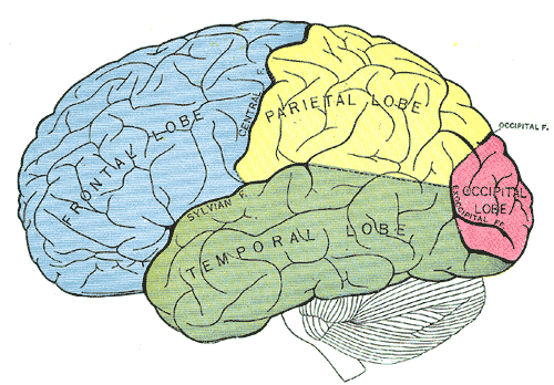
Labels on the lateral (lobes) view:
- Parietal lobe
- Central sulcus
- Parieto-occipital sulcus
- Frontal lobe
- Occipital lobe
- Lateral sulcus
- Temporal lobe
On this page
What this brain anatomy quiz covers
The quiz has two views, each on its own tab:
- Lateral (lobes): the outer surface of the left hemisphere with the four lobes shaded in different colors. You label the frontal, parietal, temporal and occipital lobes and the three grooves that separate them: the central sulcus, the lateral sulcus and the parieto-occipital sulcus.
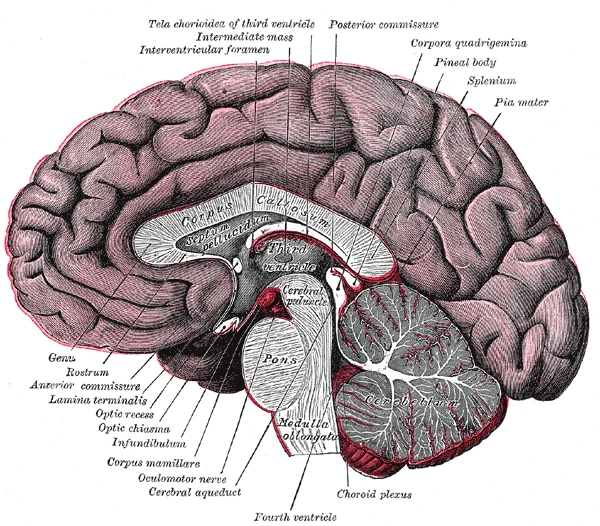
- Midsagittal section: the brain cut down the middle from front to back. This view shows what lies along the midline: the corpus callosum and septum pellucidum, the third and fourth ventricles with the cerebral aqueduct between them, the parts of the brainstem, the cerebellum, and small structures on the underside such as the optic chiasm, the infundibulum and the mammillary bodies.
That makes 23 labels in total. The 1918 plates use some names that have since changed, such as Sylvian fissure and optic chiasma; the quiz asks for the current name and Type it accepts the old one. Other printed labels on the midline plate that are not quiz answers, like genu, splenium or choroid plexus, stay visible as extra context.
- Parietal lobe
- Lies behind the central sulcus and above the lateral sulcus. It receives touch, pain and body-position signals and helps you judge space, size and distance. (lateral (lobes) view)
- Central sulcus
- The groove that separates the frontal lobe from the parietal lobe. The primary motor cortex lies just in front of it and the primary sensory cortex just behind it. (lateral (lobes) view)
- Parieto-occipital sulcus
- The groove that marks the border between the parietal and occipital lobes. It is seen mainly on the inner surface of the hemisphere and only notches the outer surface. (lateral (lobes) view)
- Frontal lobe
- The front part of each cerebral hemisphere, ahead of the central sulcus. It plans and controls voluntary movement and handles reasoning, decision-making and, usually on the left side, the production of speech. (lateral (lobes) view)
- Occipital lobe
- The back part of the hemisphere. It holds the primary visual cortex, which receives the signals that come from the eyes. (lateral (lobes) view)
- Lateral sulcus
- The deep groove on the side of the brain that separates the temporal lobe from the frontal and parietal lobes; often called the Sylvian fissure. (lateral (lobes) view)
- Temporal lobe
- Lies below the lateral sulcus. It contains the primary hearing area and, on its inner side, structures that are important for memory. (lateral (lobes) view)
- Corpora quadrigemina
- Four small rounded bumps on the back of the midbrain: the two superior colliculi, involved in visual reflexes, and the two inferior colliculi, which relay hearing. (midsagittal section view)
- Interventricular foramen
- The small opening that connects each lateral ventricle with the third ventricle; also called the foramen of Monro. (midsagittal section view)
- Pineal gland
- A small gland behind the third ventricle that releases melatonin, a hormone linked to the sleep–wake cycle. (midsagittal section view)
- Corpus callosum
- The large band of nerve fibers that connects the left and right cerebral hemispheres. In a midline cut it is the white arch above the lateral ventricle. (midsagittal section view)
- Septum pellucidum
- A thin double membrane in the midline that separates the left and right lateral ventricles, stretched between the corpus callosum and the fornix. (midsagittal section view)
- Third ventricle
- A narrow, fluid-filled space in the midline between the two halves of the thalamus. It connects with the lateral ventricles and, through the cerebral aqueduct, with the fourth ventricle. (midsagittal section view)
- Cerebral peduncle
- One of the paired stalks on the front of the midbrain, made of nerve fibers that run between the cerebrum and the brainstem and spinal cord. (midsagittal section view)
- Pons
- The rounded middle part of the brainstem, between the midbrain and the medulla oblongata. It relays signals to the cerebellum and helps regulate breathing. (midsagittal section view)
- Anterior commissure
- A small bundle of nerve fibers that crosses the midline in front of the third ventricle and links parts of the two temporal lobes. (midsagittal section view)
- Cerebellum
- The “little brain” at the back, below the occipital lobes. It coordinates movement, balance and posture; in a cut, its white matter branches like a tree (the arbor vitae). (midsagittal section view)
- Medulla oblongata
- The lowest part of the brainstem, continuous with the spinal cord. It contains centers that control breathing, heart rate and blood pressure. (midsagittal section view)
- Optic chiasm
- Where the two optic nerves meet below the brain and the fibers from the inner half of each retina cross to the opposite side. (midsagittal section view)
- Infundibulum
- The stalk that connects the hypothalamus to the pituitary gland. (midsagittal section view)
- Mammillary body
- One of a pair of small round bumps on the underside of the hypothalamus, involved in memory. (midsagittal section view)
- Cerebral aqueduct
- The narrow channel through the midbrain that carries cerebrospinal fluid from the third to the fourth ventricle. (midsagittal section view)
- Fourth ventricle
- The fluid-filled space between the pons and medulla in front and the cerebellum behind. It drains cerebrospinal fluid into the space around the brain and spinal cord. (midsagittal section view)
How to memorize the parts of the brain
- Name the lobes after the bones. Each lobe sits under the skull bone with the same name: frontal under the forehead, parietal under the crown, temporal at the temple, occipital at the back. If you have done the skull labeling quiz, you already know the map.
- Use the two big grooves as borders. The central sulcus runs from the top of the head down toward the ear and splits frontal from parietal. The lateral sulcus runs back from the front and sets the temporal lobe apart below it. Everything else falls into place around these two lines.
- Link each lobe to one job. Frontal: movement and planning. Parietal: touch and body sense. Temporal: hearing and memory. Occipital: vision. One word per lobe is enough to answer most function questions.
- Read the midline from top to bottom. Corpus callosum, septum pellucidum, third ventricle, midbrain with its aqueduct, pons, medulla. The cerebellum and fourth ventricle sit behind the pons and medulla.
- Follow the fluid. Cerebrospinal fluid flows from the lateral ventricles through the interventricular foramen into the third ventricle, down the cerebral aqueduct and into the fourth ventricle. Tracing that route ties five labels together.
Common mistakes
- Mixing up the central and lateral sulci. The central sulcus runs roughly from top to bottom; the lateral sulcus runs roughly from front to back, lower on the side of the brain.
- Putting the cerebellum in the occipital lobe. The occipital lobe is part of the cerebrum; the cerebellum is the separate, finely folded structure tucked underneath it.
- Third versus fourth ventricle. The third ventricle is higher, in the middle of the brain between the thalami; the fourth lies lower, in front of the cerebellum.
- Pons versus medulla oblongata. The pons is the rounded bulge; the medulla is the narrower part below it that continues into the spinal cord.
Keep going
The cranial nerves leave from the underside of the brain and brainstem. See where each of the twelve emerges in the cranial nerves quiz.
Printable worksheets
Prefer paper? Each of the 2 views of this quiz comes as a free one-page PDF worksheet: the same plate with every label hidden under a blank numbered box, and a numbered line to write each name. A matching answer key lists the names with the same numbers as the quiz above.
How to use it: study the plate on screen first, then print the worksheet and fill in every line from memory. Mark it with the answer key and circle the ones you missed. A day or two later, print a fresh copy and do only the circled numbers, or switch to Type it above, which asks for the names in the same order.
- Lateral (lobes) view: Worksheet · A4 · Answer key · A4
- Midsagittal section view: Worksheet · A4 · Answer key · A4
Frequently asked questions
- What are the four lobes of the brain?
- Each cerebral hemisphere has a frontal, parietal, temporal and occipital lobe, named after the skull bones that cover them. Some textbooks add a fifth region, the insula, hidden deep inside the lateral sulcus, and the limbic lobe on the inner surface.
- What is the difference between a sulcus and a fissure?
- Both are grooves on the surface of the brain. Fissure is usually kept for the deepest ones, such as the longitudinal fissure between the hemispheres; sulcus is used for the rest. Older books, including the 1918 plates in this quiz, called the central and lateral sulci fissures.
- What are the three parts of the brainstem?
- From top to bottom: the midbrain, the pons and the medulla oblongata. In the midline section of this quiz you can see all three, with the cerebral peduncle and corpora quadrigemina belonging to the midbrain.
- Where is the cerebrospinal fluid in a midline section?
- In the third ventricle between the two halves of the thalamus, in the cerebral aqueduct that runs through the midbrain and in the fourth ventricle between the brainstem and the cerebellum. The lateral ventricles lie to each side of the midline, beneath the corpus callosum.
- Is the cerebellum part of the brainstem?
- No. The cerebellum sits behind the brainstem and is attached to it by three pairs of stalks, but it is a separate part of the brain that coordinates movement and balance.
Sources
- Henry Gray, Anatomy of the Human Body, 20th ed., ed. W. H. Lewis (Philadelphia: Lea & Febiger, 1918), Neurology: The Brain, Figs. 720 and 728
- Federative International Programme for Anatomical Terminology (FIPAT), Terminologia Anatomica, 2nd ed. (2019), TA2 Viewer
- OpenStax, Anatomy and Physiology 2e, section 13.2: The Central Nervous System
Image credits
- Lateral (lobes) view: Illustration: Henry Gray, Anatomy of the Human Body (1918), public domain, via Wikimedia Commons.
- Midsagittal section view: Illustration: Henry Gray, Anatomy of the Human Body (1918), public domain, via Wikimedia Commons.
Licenses, sources and changes for every illustration are listed on the image credits page.