
Skull Labeling Quiz: Bones, Sutures and Foramina
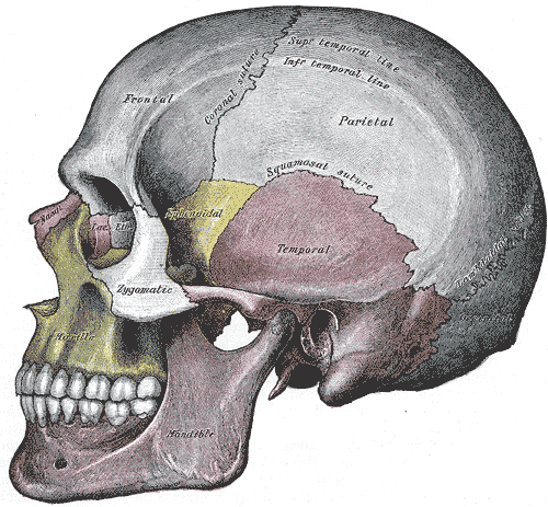
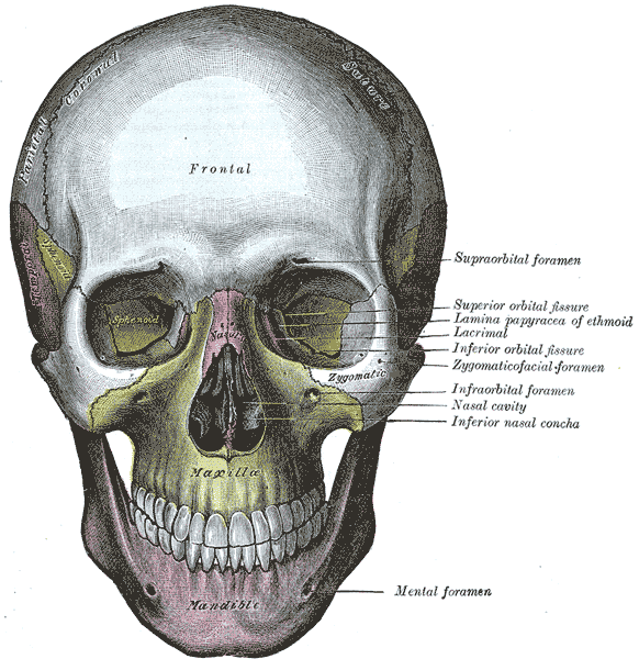
This skull labeling quiz covers the bones, sutures and openings of the human skull in side and front views, drawn from Henry Gray's Anatomy of the Human Body (1918).


Study
Labels on the lateral view:
- Superior temporal line
- Inferior temporal line
- Coronal suture
- Frontal bone
- Parietal bone
- Squamous suture
- Sphenoid bone
- Nasal bone
- Lacrimal bone
- Ethmoid bone
- Temporal bone
- Lambdoid suture
- Zygomatic bone
- Occipital bone
- Maxilla
- Mandible
On this page
How the skull labeling quiz works
The quiz shows two classic engravings of the skull: a side (lateral) view and a front (anterior) view. Switch between them with the tabs above the plate. Each view has its own set of labels, 33 in total covering 25 different bones, sutures, openings and landmarks, so you can master one view before moving on.
Every view works in five modes. Study shows all the names; tap a label to read what the structure is and why it matters. Label it hides the names and gives you a word bank to place. Find it names a structure and asks you to tap its box; the prompt stays until you find it, but only your first tap counts toward the score. Type it asks you to write each name from memory; capital letters and accents are ignored, accepted alternative names count, and a single wrong letter is forgiven with a spelling note. Timed is Find it against the clock, and your best time is saved in this browser. After every round you get your score, a list of your mistakes with the right answers, and a button to retry only the ones you missed.
What the quiz covers
The lateral view focuses on the main cranial and facial bones, the three large sutures you can see from the side and the two temporal lines. The anterior view adds the bones around the orbit and nose and the openings on the face that nerves and vessels pass through. Some printed labels on the plates are not part of the quiz, such as the muscle names; they are left visible when they use current terms.
- Superior temporal line
- The upper of two curved ridges on the frontal and parietal bones. The temporal fascia, which covers the temporalis muscle, attaches along it. (lateral view)
- Inferior temporal line
- The lower temporal line. It marks the upper edge of the area where the temporalis muscle arises. (lateral view)
- Coronal suture
- The joint between the frontal bone and the two parietal bones, running across the top of the skull from side to side.
- Frontal bone
- Forms the forehead and the roofs of the orbits. It meets the two parietal bones at the coronal suture.
- Parietal bone
- One of a pair of curved bones that form most of the roof and upper sides of the cranium. The two parietals meet in the midline at the sagittal suture. (lateral view)
- Squamous suture
- The curved, overlapping joint between the squamous part of the temporal bone and the parietal bone. (lateral view)
- Sphenoid bone
- A bat-shaped bone in the middle of the skull base. From the side only its greater wing shows, between the frontal and temporal bones; inside the orbit it forms part of the back wall.
- Nasal bone
- One of two small, oblong bones that meet in the midline to form the bridge of the nose.
- Lacrimal bone
- The smallest facial bone, at the front of the medial wall of the orbit. It helps form the fossa for the lacrimal sac, which drains tears into the nose.
- Ethmoid bone
- A light, honeycombed bone between the orbits. Its thin orbital plate forms most of the medial wall of the orbit, and its upper part forms the roof of the nasal cavity. (lateral view)
- Temporal bone
- Forms the lower side and part of the base of the skull and houses the middle and inner ear. The thin, flat part seen on the side of the head is its squamous part. (lateral view)
- Lambdoid suture
- The joint between the occipital bone and the two parietal bones at the back of the skull, shaped like the Greek letter lambda (λ). (lateral view)
- Zygomatic bone
- The cheekbone. Its temporal process joins the zygomatic process of the temporal bone to form the zygomatic arch.
- Occipital bone
- Forms the back and much of the base of the skull. The foramen magnum, where the brainstem continues into the spinal cord, passes through it. (lateral view)
- Maxilla
- The upper jaw. The two maxillae hold the upper teeth and form most of the hard palate, the floor of each orbit and the sides of the nasal cavity.
- Mandible
- The lower jaw and the largest facial bone. It is the only skull bone joined to the cranium by a freely movable joint, the temporomandibular joint.
- Supraorbital foramen
- An opening, or sometimes only a notch, in the upper rim of the orbit. The supraorbital nerve and vessels pass through it to the forehead. (anterior view)
- Superior orbital fissure
- A slit between the greater and lesser wings of the sphenoid at the back of the orbit. The oculomotor, trochlear, ophthalmic and abducens nerves enter the orbit through it. (anterior view)
- Orbital plate of ethmoid
- The paper-thin plate of the ethmoid bone that forms most of the medial wall of the orbit. Its old name, lamina papyracea, means “papery layer”. (anterior view)
- Inferior orbital fissure
- A gap in the floor of the orbit between the maxilla and the greater wing of the sphenoid. The infraorbital and zygomatic nerves pass through it. (anterior view)
- Zygomaticofacial foramen
- A small opening on the outer surface of the zygomatic bone for the zygomaticofacial nerve and vessels. (anterior view)
- Infraorbital foramen
- An opening in the maxilla just below the orbit. The infraorbital nerve and vessels come out through it onto the face. (anterior view)
- Nasal cavity
- The air passage behind the nose, divided into right and left halves by the nasal septum. (anterior view)
- Inferior nasal concha
- A separate, scroll-shaped bone on the side wall of each nasal cavity. The superior and middle conchae above it are parts of the ethmoid bone. (anterior view)
- Mental foramen
- An opening on the outer surface of the mandible, below the premolar teeth, for the mental nerve and vessels. (anterior view)
Tips for memorizing the bones of the skull
- Sort the bones into two families first. Eight cranial bones form the case around the brain: frontal, two parietal, two temporal, occipital, sphenoid and ethmoid. A common memory aid is “Old People From Texas Eat Spiders” (occipital, parietal, frontal, temporal, ethmoid, sphenoid). The fourteen facial bones are easier once you know “Virgil Can Not Make My Pet Zebra Laugh” (vomer, conchae, nasal, maxilla, mandible, palatine, zygomatic, lacrimal).
- Follow the sutures like borders on a map. The coronal suture runs across the top of the head like the band of a crown, the lambdoid suture forms an upside-down V at the back, and the squamous suture arches over the ear. If you know which bones each suture separates, the bones fall into place.
- Use the orbit as a checkpoint. Seven bones build each orbit: frontal, sphenoid, zygomatic, maxilla, ethmoid, lacrimal and palatine. Naming them in order around the rim and walls is a quick self-test for both views.
- Learn openings by what passes through them. The superior orbital fissure lets the nerves that move the eye enter the orbit, the supraorbital and infraorbital foramina let sensory nerves reach the skin of the forehead and cheek, and the mental foramen does the same for the chin. Linking each opening to its contents prepares you for the cranial nerves quiz.
- Space your practice. Do one view in Label it today, the same view in Type it tomorrow, then move to Timed. Retrying only your mistakes at the end of each round is the fastest way to close the gaps.
Common mistakes to watch for
- Zygomatic bone versus zygomatic arch. The arch is formed by two bones: the temporal process of the zygomatic bone and the zygomatic process of the temporal bone. The label on the cheek is the bone itself.
- Sphenoid on the side of the head. Many students expect the sphenoid only in the middle of the skull base, but its greater wing reaches the side of the skull just in front of the temporal bone.
- Lacrimal versus ethmoid in the orbit. The lacrimal bone is the small piece at the front of the medial wall, next to the nose; the larger, paper-thin plate behind it belongs to the ethmoid.
- Superior versus inferior orbital fissure. The superior one sits between the two wings of the sphenoid, high in the back of the orbit; the inferior one runs along the floor, between the sphenoid and the maxilla.
- Inferior nasal concha. It is a bone of its own. The superior and middle conchae, higher up, are parts of the ethmoid.
Keep going
Once the skull feels solid, put it in context with the rest of the trunk skeleton in the axial skeleton quiz, or test the whole body in the bones quiz.
Printable worksheets
Prefer paper? Each of the 2 views of this quiz comes as a free one-page PDF worksheet: the same plate with every label hidden under a blank numbered box, and a numbered line to write each name. A matching answer key lists the names with the same numbers as the quiz above.
How to use it: study the plate on screen first, then print the worksheet and fill in every line from memory. Mark it with the answer key and circle the ones you missed. A day or two later, print a fresh copy and do only the circled numbers, or switch to Type it above, which asks for the names in the same order.
- Lateral view: Worksheet · A4 · Answer key · A4
- Anterior view: Worksheet · A4 · Answer key · A4
Frequently asked questions
- How many bones are in the human skull?
- The adult skull has 22 bones: 8 cranial bones that enclose the brain and 14 facial bones. That count leaves out the six tiny ear ossicles inside the temporal bones and the hyoid bone in the neck.
- Which skull bones come in pairs?
- The paired bones are the parietal and temporal bones of the cranium, and the maxillae, zygomatic, nasal, lacrimal, palatine and inferior nasal conchae of the face. The frontal, occipital, sphenoid, ethmoid, vomer and mandible are single bones in the midline.
- Is the hyoid bone part of the skull?
- No. The hyoid sits in the upper neck and does not form a joint with any other bone; muscles and ligaments hold it in place, including a ligament that runs to the styloid process of each temporal bone.
- What is the pterion, and why do anatomy courses stress it?
- The pterion is the small area on the side of the skull where the frontal, parietal, temporal and sphenoid bones meet. The bone there is thin and the middle meningeal artery runs just inside it, so a blow to this spot can tear the artery and cause bleeding between the skull and the dura.
- Why do some printed labels on the plates differ from the answer names?
- The illustrations were published in 1918 and use the terms of that time, such as squamosal suture, lambdoidal suture and lamina papyracea. The quiz uses current names, and Type it also accepts the older names you may still see in some textbooks.
- What is the difference between a foramen and a fissure?
- A foramen is a round or oval hole, such as the supraorbital or infraorbital foramen. A fissure is a narrow slit between two bones or two parts of a bone, such as the superior and inferior orbital fissures at the back of the orbit.
Sources
- Henry Gray, Anatomy of the Human Body, 20th ed., ed. W. H. Lewis (Philadelphia: Lea & Febiger, 1918), Osteology: The Skull, Figs. 188 and 190
- Federative International Programme for Anatomical Terminology (FIPAT), Terminologia Anatomica, 2nd ed. (2019), TA2 Viewer
- OpenStax, Anatomy and Physiology 2e, section 7.2: The Skull
Image credits
- Lateral view: Illustration: Henry Gray, Anatomy of the Human Body (1918), public domain, via Wikimedia Commons.
- Anterior view: Illustration: Henry Gray, Anatomy of the Human Body (1918), public domain, via Wikimedia Commons.
Licenses, sources and changes for every illustration are listed on the image credits page.