
Axial Skeleton Quiz: Skull, Vertebral Column and Thorax
This axial skeleton quiz covers the bones along the midline of the body, the skull, the vertebral column, the sternum and the ribs, using three plates from Henry Gray's Anatomy of the Human Body (1918).



Study
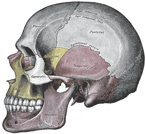
Labels on the skull view:
- Frontal bone
- Parietal bone
- Sphenoid bone
- Nasal bone
- Lacrimal bone
- Ethmoid bone
- Temporal bone
- Zygomatic bone
- Occipital bone
- Maxilla
- Mandible
On this page
What this axial skeleton quiz covers
The skeleton is usually split in two. The axial skeleton is the column of bones along the midline that protects the brain, the spinal cord and the organs of the chest; the appendicular skeleton is everything that hangs from it, the limbs and the girdles that attach them. This quiz covers the axial part in three views, each on its own tab:
- Skull: the side view of the skull with its eleven bones, from the frontal bone at the front to the occipital bone at the back and the mandible below. Sutures and temporal lines are printed on the plate and stay visible as extra help.
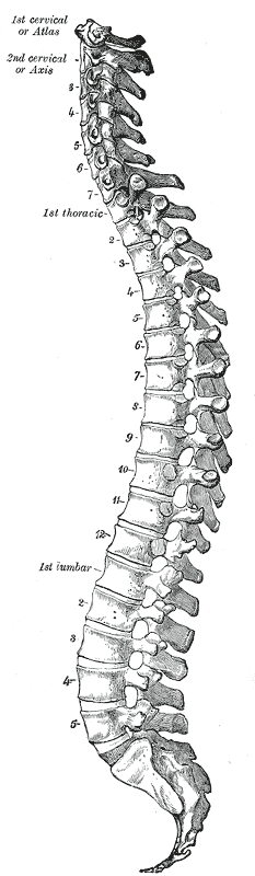
- Vertebral column: the whole column seen from the left side, with the atlas and axis at the top, one box in each of the cervical, thoracic and lumbar regions, the vertebra prominens, an intervertebral disc, and the sacrum and coccyx at the bottom. Gray numbers each vertebra within its region; those numbers stay visible.
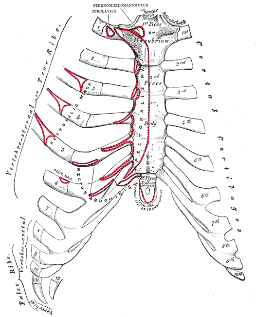
- Sternum and ribs: the front of the chest, with the three parts of the sternum, the jugular notch and the sternal angle, and the groups of ribs: true, false and floating.
That is 28 labels in all. Some printed labels use 1918 wording, such as "2nd piece or body" for the body of the sternum and vertebral ribs for the floating ribs; the quiz asks for the current name. For the regions of the column, the box sits on one vertebra of the region, as the description of each label explains.
- Frontal bone
- Forms the forehead and the roofs of the orbits. It meets the two parietal bones at the coronal suture. (skull view)
- Parietal bone
- One of a pair of curved bones that form most of the roof and upper sides of the cranium. The two parietals meet in the midline at the sagittal suture. (skull view)
- Sphenoid bone
- A bat-shaped bone in the middle of the skull base. From the side only its greater wing shows, between the frontal and temporal bones; inside the orbit it forms part of the back wall. (skull view)
- Nasal bone
- One of two small, oblong bones that meet in the midline to form the bridge of the nose. (skull view)
- Lacrimal bone
- The smallest facial bone, at the front of the medial wall of the orbit. It helps form the fossa for the lacrimal sac, which drains tears into the nose. (skull view)
- Ethmoid bone
- A light, honeycombed bone between the orbits. Its thin orbital plate forms most of the medial wall of the orbit, and its upper part forms the roof of the nasal cavity. (skull view)
- Temporal bone
- Forms the lower side and part of the base of the skull and houses the middle and inner ear. The thin, flat part seen on the side of the head is its squamous part. (skull view)
- Zygomatic bone
- The cheekbone. Its temporal process joins the zygomatic process of the temporal bone to form the zygomatic arch. (skull view)
- Occipital bone
- Forms the back and much of the base of the skull. The foramen magnum, where the brainstem continues into the spinal cord, passes through it. (skull view)
- Maxilla
- The upper jaw. The two maxillae hold the upper teeth and form most of the hard palate, the floor of each orbit and the sides of the nasal cavity. (skull view)
- Mandible
- The lower jaw and the largest facial bone. It is the only skull bone joined to the cranium by a freely movable joint, the temporomandibular joint. (skull view)
- Atlas
- The first cervical vertebra (C1), a ring of bone with no body that carries the skull. Nodding the head “yes” happens between the atlas and the skull. (vertebral column view)
- Axis
- The second cervical vertebra (C2). Its tooth-like dens sticks up into the ring of the atlas, the pivot for turning the head “no”. (vertebral column view)
- Cervical vertebrae
- The seven vertebrae of the neck (C1–C7). They are the smallest vertebrae and each transverse process has a hole for the vertebral artery. Box on the fifth (C5). (vertebral column view)
- Vertebra prominens
- The seventh cervical vertebra (C7). Its long spinous process is the bump you feel at the base of the back of the neck, a useful starting point for counting vertebrae. (vertebral column view)
- Thoracic vertebrae
- The twelve vertebrae of the chest (T1–T12). Each one joins a pair of ribs. Box on the sixth (T6). (vertebral column view)
- Lumbar vertebrae
- The five vertebrae of the lower back (L1–L5). They have the largest bodies because they carry the most weight. Box on the third (L3). (vertebral column view)
- Intervertebral disc
- The pad of fibrocartilage between the bodies of two vertebrae. It absorbs shock and lets the spine bend. Box on the disc between L3 and L4. (vertebral column view)
- Sacrum
- A large triangular bone made of five fused sacral vertebrae. It joins the hip bones at the sacroiliac joints and forms the back wall of the pelvis. (vertebral column view)
- Coccyx
- The tailbone: three to five small fused vertebrae at the lower end of the column. (vertebral column view)
- Jugular notch
- The dip in the top of the manubrium, between the inner ends of the clavicles. You can feel it at the base of the front of the neck. (sternum and ribs view)
- Manubrium
- The upper part of the sternum, shaped like a broad tie knot. The clavicles and the first pair of ribs join it. (sternum and ribs view)
- Sternal angle
- The slight ridge where the manubrium meets the body of the sternum. The second ribs join the sternum here, which makes it the standard landmark for counting ribs. (sternum and ribs view)
- Body of sternum
- The long middle part of the sternum. The costal cartilages of the second to seventh ribs join its edges. (sternum and ribs view)
- Xiphoid process
- The small pointed lower end of the sternum. It is cartilage in young people and slowly turns to bone with age. (sternum and ribs view)
- True ribs
- Ribs 1 to 7. Each one is joined directly to the sternum by its own costal cartilage. (sternum and ribs view)
- False ribs
- Ribs 8 to 12. They do not reach the sternum on their own: the cartilages of ribs 8 to 10 join the cartilage above, and ribs 11 and 12 end free. (sternum and ribs view)
- Floating ribs
- Ribs 11 and 12, the last two false ribs. Their front ends are free in the muscles of the abdominal wall. Gray's plate calls them vertebral ribs. (sternum and ribs view)
How to learn the axial skeleton
- Count the column at mealtimes. 7 cervical, 12 thoracic, 5 lumbar: breakfast at 7, lunch at 12 and dinner at 5. Then 5 fused sacral vertebrae and a small coccyx.
- Follow the curves. Seen from the side, the column has four curves: the neck and lower back curve forward, the chest and sacrum curve backward. The second view shows them clearly; the region boundaries sit near the points where the curve changes.
- Use landmarks you can feel. The vertebra prominens is the bump at the base of your neck, the jugular notch is the dip above your sternum and the sternal angle is the small ridge a few finger-widths below it. Rib 2 meets the sternum at the sternal angle, so you can count down the ribs from there.
- Group the ribs by how they reach the sternum. Ribs 1–7 have their own cartilage to the sternum (true). Ribs 8–10 join the cartilage above (false). Ribs 11–12 end free (false and floating).
- Revisit the skull with more detail. This quiz only asks for the bones of the skull; the skull labeling quiz adds the sutures, the temporal lines and a front view.
Common mistakes
- Atlas versus axis. The atlas (C1) comes first and carries the skull; the axis (C2) is below it and has the dens. Alphabetical order helps: A-t comes before A-x.
- Thoracic versus lumbar vertebrae. Lumbar vertebrae are much larger and have no joints for ribs. On the plate, the bodies get visibly bigger as you go down the column.
- Sacrum and coccyx as one bone. They are two separate bones joined by a small joint; the sacrum is the large triangle, the coccyx the small tail below it.
- Calling all lower ribs floating. Only ribs 11 and 12 float; ribs 8 to 10 are false but attached to the cartilage above.
Keep going
The nerves that leave the skull through its openings are covered in the cranial nerves quiz. For a broader review of how the whole skeleton is organized, try the skeletal system quiz.
Printable worksheets
Prefer paper? Each of the 3 views of this quiz comes as a free one-page PDF worksheet: the same plate with every label hidden under a blank numbered box, and a numbered line to write each name. A matching answer key lists the names with the same numbers as the quiz above.
How to use it: study the plate on screen first, then print the worksheet and fill in every line from memory. Mark it with the answer key and circle the ones you missed. A day or two later, print a fresh copy and do only the circled numbers, or switch to Type it above, which asks for the names in the same order.
- Skull view: Worksheet · A4 · Answer key · A4
- Vertebral column view: Worksheet · A4 · Answer key · A4
- Sternum and ribs view: Worksheet · A4 · Answer key · A4
Frequently asked questions
- What bones make up the axial skeleton?
- The axial skeleton is the central axis of the body: the skull, the vertebral column (including the sacrum and coccyx), the sternum and the ribs. Most textbooks also include the hyoid bone and the small bones of the middle ear. In an adult it has about 80 bones.
- How many vertebrae are there in each region?
- Seven cervical vertebrae in the neck, twelve thoracic vertebrae in the chest and five lumbar vertebrae in the lower back. Below them, five sacral vertebrae fuse into the sacrum and three to five small ones form the coccyx.
- What is the difference between true, false and floating ribs?
- The first seven pairs are true ribs: each joins the sternum through its own costal cartilage. Pairs 8 to 12 are false ribs because they do not reach the sternum directly. The last two pairs, 11 and 12, are also called floating ribs because their front ends are free.
- Why are the atlas and axis named that way?
- The atlas, the first cervical vertebra, holds up the skull the way the Titan Atlas held up the sky in Greek myth. The axis, the second, has a peg (the dens) that acts as the pivot around which the atlas and skull turn.
- What is the sternal angle used for?
- It is a palpable ridge where the manubrium meets the body of the sternum, and the second rib joins the sternum at that level. Clinicians use it as a starting point to count ribs and intercostal spaces, for example when listening to the heart.
Sources
- Henry Gray, Anatomy of the Human Body, 20th ed., ed. W. H. Lewis (Philadelphia: Lea & Febiger, 1918), Osteology, Figs. 111, 115 and 188
- Federative International Programme for Anatomical Terminology (FIPAT), Terminologia Anatomica, 2nd ed. (2019), TA2 Viewer
- OpenStax, Anatomy and Physiology 2e, chapter 7: Axial Skeleton
Image credits
- Skull view: Illustration: Henry Gray, Anatomy of the Human Body (1918), public domain, via Wikimedia Commons.
- Vertebral column view: Illustration: Henry Gray, Anatomy of the Human Body (1918), public domain, via Wikimedia Commons.
- Sternum and ribs view: Illustration: Henry Gray, Anatomy of the Human Body (1918), public domain, via Wikimedia Commons.
Licenses, sources and changes for every illustration are listed on the image credits page.