
Bones Quiz: Label the Major Bones of the Skeleton
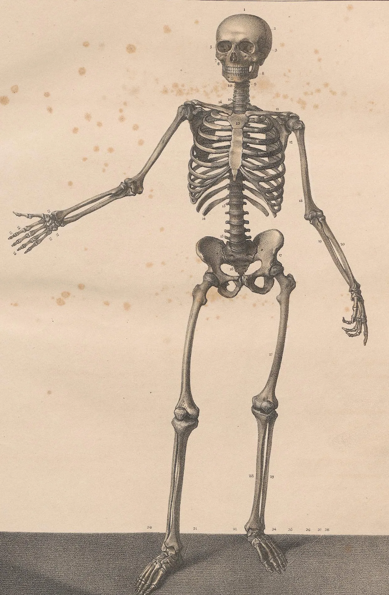
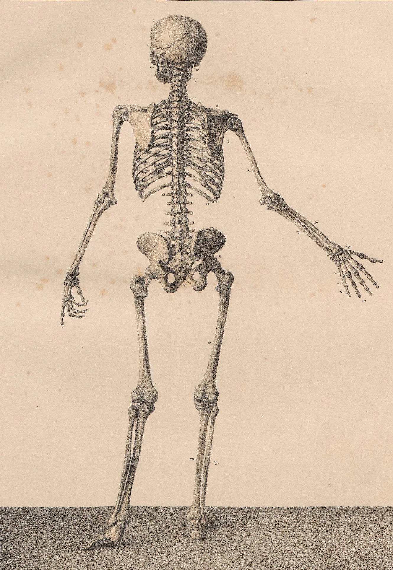
This bones quiz asks you to label the major bones of the human skeleton, from the skull to the feet, on a front and a back view of a complete skeleton from Bourgery's anatomical atlas (1831).
Study
Labels on the anterior (front) view:
- Frontal bone
- Parietal bone
- Nasal bone
- Temporal bone
- Zygomatic bone
- Maxilla
- Mandible
- Cervical vertebrae
- Clavicle
- Sternum
- Scapula
- Humerus
- Lumbar vertebrae
- Carpal bones
- Ribs
- Phalanges of the hand
- Metacarpals
- Ulna
- Radius
- Hip bone
- Sacrum
- Femur
- Patella
- Tibia
- Fibula
- Talus
- Calcaneus
- Metatarsals
On this page
What this bones quiz covers
This quiz is about the big picture: finding the major bones of the whole skeleton, from the skull to the feet, on two full-length drawings. Switch between them with the tabs above the plate:
- Anterior (front): the skeleton seen from the front with one arm held out to the side, so the bones of the forearm and hand are easy to see. You label the bones of the skull and face, the cervical and lumbar vertebrae, the clavicle, sternum, ribs and scapula, every bone of the arm and leg, the kneecap and the main bones of the foot.
- Posterior (back): the same skeleton from behind. Here you find the occipital bone, the thoracic vertebrae, the sacrum and hip bones from the back, and the long bones of the limbs again, this time seen from the other side.
There are 49 labels in all. The plates come from Bourgery and Jacob's anatomical atlas of 1831. Bourgery marks the bones with small numbers and names them on a separate legend page in French; each quiz box sits on one of those numbers. For regions of the spine he marks the first and last vertebra with the same number, so those boxes span or sit at the end of the region, as the description of each label explains. The other numbers stay visible: without the legend they do not name anything.
- Frontal bone
- The forehead bone. It forms the front of the skull and the roofs of the eye sockets. (anterior (front) view)
- Parietal bone
- One of the two bones that form the top and upper sides of the skull.
- Nasal bone
- One of the two small bones that form the bridge of the nose. (anterior (front) view)
- Temporal bone
- The bone at the side of the skull around the ear. It holds the hearing and balance organs.
- Zygomatic bone
- The cheekbone, below and to the side of the eye socket. (anterior (front) view)
- Maxilla
- The upper jaw. The two maxillae hold the upper teeth and form part of the eye sockets and the hard palate. (anterior (front) view)
- Mandible
- The lower jaw, the only movable bone of the skull. It holds the lower teeth. (anterior (front) view)
- Cervical vertebrae
- The seven vertebrae of the neck. Bourgery marks the region with a 10 at each end.
- Clavicle
- The collarbone, between the sternum and the shoulder blade.
- Sternum
- The breastbone, in the middle of the chest. The costal cartilages of the upper ribs join it. (anterior (front) view)
- Scapula
- The shoulder blade, a flat triangular bone on the back of the ribs.
- Humerus
- The bone of the upper arm, from the shoulder to the elbow.
- Lumbar vertebrae
- The five large vertebrae of the lower back. They have the largest bodies of the whole column because they carry the most weight.
- Carpal bones
- The eight small bones of the wrist, in two rows.
- Ribs
- Twelve pairs of curved bones that form the cage around the chest.
- Phalanges of the hand
- The bones of the fingers: three in each finger and two in the thumb.
- Metacarpals
- The five long bones of the palm.
- Ulna
- The forearm bone on the little-finger side.
- Radius
- The forearm bone on the thumb side.
- Hip bone
- Each of the two large bones of the pelvis, formed by the fused ilium, ischium and pubis.
- Sacrum
- The triangular bone at the base of the spine, made of five fused vertebrae; the coccyx hangs below it.
- Femur
- The thigh bone, the longest and strongest bone of the body.
- Patella
- The kneecap, a small bone inside the tendon of the quadriceps in front of the knee. (anterior (front) view)
- Tibia
- The shinbone: the larger, inner bone of the leg.
- Fibula
- The thin outer bone of the leg.
- Talus
- The ankle bone, which carries the weight of the body from the tibia to the foot. (anterior (front) view)
- Calcaneus
- The heel bone, the largest bone of the foot.
- Metatarsals
- The five long bones in the middle of the foot. (anterior (front) view)
- Occipital bone
- The bone at the back and base of the skull, with the large opening for the spinal cord. (posterior (back) view)
- Thoracic vertebrae
- The twelve vertebrae of the chest, each joined to a pair of ribs. (posterior (back) view)
How to learn the bones of the skeleton
- Split the skeleton in two. The axial skeleton runs down the middle: skull, vertebral column, sternum and ribs. The appendicular skeleton is everything attached to it: the shoulder and hip girdles and the limbs. Learn one half at a time.
- Go from top to bottom. Skull, neck, chest, spine, pelvis, then each limb from the shoulder or hip down to the fingers or toes. Saying the bones in order turns the skeleton into a route instead of a list.
- Use the parallels between arm and leg. One bone in the upper segment (humerus, femur), two in the lower segment (radius and ulna, tibia and fibula), then a group of small bones, five long bones and the phalanges. The patella has no equivalent at the elbow, which makes it easy to remember.
- Feel the bones on yourself. You can feel your clavicle, the edge of your scapula, your sternum, the iliac crest of your hip bone, your patella and the front of your tibia. Touching a bone while saying its name helps it stick.
- Then zoom in. Once you know where each bone is, the axial skeleton quiz and the appendicular skeleton quiz go into the regions in more detail, on larger plates.
Common mistakes
- Radius and ulna. With the palm facing forward, the radius is on the thumb side and the ulna on the little-finger side. In the front view the outstretched arm makes this easy to check.
- Tibia and fibula. The tibia is the thick inner bone; the fibula is the thin outer one.
- Sacrum and hip bones. The sacrum is the triangular bone at the back of the pelvis, part of the spine; the two large hip bones form the sides and front.
- Scapula from the front. In the front view the scapula is mostly hidden behind the ribs; only its upper edge and the shoulder joint show. The back view shows it clearly.
Keep going
For the functions of the skeleton, the types of bone and the parts of a long bone, try the skeletal system quiz. To learn the bumps, holes and grooves on individual bones, go on to the bone markings quiz.
Printable worksheets
Prefer paper? Each of the 2 views of this quiz comes as a free one-page PDF worksheet: the same plate with every label hidden under a blank numbered box, and a numbered line to write each name. A matching answer key lists the names with the same numbers as the quiz above.
How to use it: study the plate on screen first, then print the worksheet and fill in every line from memory. Mark it with the answer key and circle the ones you missed. A day or two later, print a fresh copy and do only the circled numbers, or switch to Type it above, which asks for the names in the same order.
- Anterior (front) view: Worksheet · A4 · Answer key · A4
- Posterior (back) view: Worksheet · A4 · Answer key · A4
Frequently asked questions
- How many bones are in the human body?
- An adult skeleton usually has 206 bones. A newborn has around 270 bony pieces; many of them fuse during childhood, for example the bones of the skull, the sacrum and the hip bones. The exact count varies a little from person to person because of small extra bones such as sesamoid bones.
- What is the longest bone in the body?
- The femur, or thigh bone. It is also one of the strongest. The smallest bone is the stapes, one of the three tiny bones of the middle ear, which are too small to show on a whole-skeleton plate.
- What is the difference between the tibia and the fibula?
- Both are in the lower leg. The tibia, or shinbone, is the thick bone on the inner side that carries the weight of the body. The fibula is the thin bone on the outer side; it carries little weight but anchors muscles and forms the outer bump of the ankle.
- Which bones make up the skull?
- The skull has 22 bones. Eight form the braincase (frontal, two parietal, two temporal, occipital, sphenoid and ethmoid) and fourteen form the face, including the maxillae, the zygomatic bones, the nasal bones and the mandible. This quiz asks for the main ones you can see from the front and back.
- Why does the quiz use an 1831 atlas?
- Full-length skeleton drawings are not part of Gray's 1918 plates, and good public-domain whole-body drawings are rare. Bourgery's atlas, engraved by Nicolas-Henri Jacob, is one of the most accurate of its time, and the Heidelberg University Library scan is detailed enough to label. The names in the quiz are the current English ones, not the 1831 French ones.
Sources
- Jean-Baptiste Marc Bourgery and Nicolas-Henri Jacob, Traité complet de l'anatomie de l'homme, Atlas I (Paris, 1831), Pl. 2 and 3 with their legend; digitization: Heidelberg University Library
- Federative International Programme for Anatomical Terminology (FIPAT), Terminologia Anatomica, 2nd ed. (2019), TA2 Viewer
- OpenStax, Anatomy and Physiology 2e, section 7.1: Divisions of the Skeletal System
Image credits
- Anterior (front) view: Illustration: J.-M. Bourgery and N.-H. Jacob, Traité complet de l'anatomie de l'homme (1831–1854), public domain, via Wikimedia Commons. Digitization: Heidelberg University Library.
- Posterior (back) view: Illustration: J.-M. Bourgery and N.-H. Jacob, Traité complet de l'anatomie de l'homme (1831–1854), public domain, via Wikimedia Commons. Digitization: Heidelberg University Library.
Licenses, sources and changes for every illustration are listed on the image credits page.